

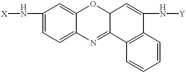
Amino acid substituted-cresyl violet, synthetic fluorogenic substrates for the analysis of agents in individual in vivo cells or tissue
a technology of in vivo cells and synthetic fluorogenic substrates, which is applied in the direction of fluorescence/phosphorescence, instruments, organic chemistry, etc., can solve the imbalance of positive and negative regulation, tumor invasion and metastasis, and ineffective defense of kupffer cells and pit cells
- Summary
- Abstract
- Description
- Claims
- Application Information
AI Technical Summary
Problems solved by technology
Method used
Image


Examples
example 1
Synthesis of [Ala-Pro].sup.2 -cresyl violet
Z-alanyl-proline dicyclohexylamine salt (5 g, 10 mmole) was suspended in 80 ml dimethylformamide / pyridine (1:1 v / v) and cooled to 0.degree. C. Then, 1-(3-methylaminopropyl)-3-ethylcarbodiimide hydrochloride (2 g, 10.4 mmole) was added. After 20 min at 0.degree. C., cresyl violet hydrochloride (1 g, 3.2 mmole) was added. The reaction mixture was allowed to stir 18 h while the temperature was raised slowly to room temp. The solvents were removed at 50.degree. C. and the residue was dissolved in 300 ml ethyl acetate. The solution was washed twice with 100 ml 1 N aqueous hydrochloric acid, once with 50 ml saturated aqueous brine, twice with 100 ml saturated aqueous sodium bicarbonate, and once with 100 ml saturated aqueous brine. The ethyl acetate solution was dried over anhydrous magnesium sulfate, filtered, stripped on a rotary evaporator and dried overnight under high vacuum. The crude product (1.5 g) was chromatographed on silica gel using ...
example 2
Isolation of Hepatocytes
Hepatocytes were isolated by collagenase perfusion of livers of male Wistar rats (200-250 g; HSD Animal Farm, Zeist, The Netherlands) after 24 h of starvation as described previously (30). The animals were exposed to a controlled dark-light cycle (light: 7:00 a.m.-7:00 p.m.) throughout the acclimatization period of at least 1 week. Before starvation, animals had free access to food (standard chow diet; Hope Farms, Woerden, The Netherlands) and water. The animals had always free access to water. During operation, the animals were under Nembutal anaesthesia. Animal care was performed according to the guidelines of the University of Amsterdam. Hepatocytes (5-10 mg dry mass / ml) were kept in Krebs-Henseleit bicarbonate medium containing 1.3 mM Ca.sup.2+, 10 mM sodium Hepes (pH 7.4), 20 mM glucose and 1 mM octanoate on ice until enzyme assays. Homogenates were prepared by freezing cell suspensions in liquid nitrogen and subsequent thawing. One volume of homogenates...
example 3
Analysis of DPPIV Activity
DPPIV activity was determined in hepatocytes using 6 approaches. Activity was determined in living hepatocytes with confocal scanning laser microscopy, image processing and analysis, flow cytometry, and fluorometry. Fluorometry was also used for the determination of activity in homogenates of hepatocytes and in membrane fractions of hepatocytes. Incubations were started at t=0 by adding an aliquot of 60 .mu.l hepatocytes to 3 ml Krebs-Henseleit medium containing 0-50 .mu.M (Ala-Pro).sup.2 -cresyl violet in the presence or absence of 0-50 .mu.M Ala-Pip.sup.p (OPh-4-Cl).sub.2 (Enzyme Systems Products), which is a selective DPPIV inhibitor (31A,32A). Substrate and inhibitor were dissolved first in dimethylsulfoxide. The final concentration of dimethylsulfoxide in the incubation medium was 0.5% (v / v). Incubations were carried out at 20.degree. C. Confocal scanning laser microscopy was performed after 100 .mu.l of an assay medium containing hepatocytes was broug...
PUM
| Property | Measurement | Unit |
|---|---|---|
| temperature | aaaaa | aaaaa |
| wavelengths | aaaaa | aaaaa |
| wavelengths | aaaaa | aaaaa |
Abstract
Description
Claims
Application Information