Methods and articles for regenerating bone or peridontal tissue

a technology of peridontal tissue and regenerative methods, applied in the field of regenerating bone or peridontal tissue, can solve the problems of limited amount of tissue available for transfer, morbidity at the donor site, and current therapeutic options for increasing bone or periodontal tissue mass

Inactive Publication Date: 2002-06-25
WL GORE & ASSOC INC
View PDF22 Cites 222 Cited by
  • Summary
  • Abstract
  • Description
  • Claims
  • Application Information

AI Technical Summary

Benefits of technology

Members of the TGF-Beta Superfamily of proteins (TGF-Beta Proteins) have numerous potential effects, most of which stimulate growth and development of cells and tissues. TGF-Beta proteins are placed within the space established, by implantation of the TP device, in order to stimulate cell differentiation, cell proliferation, cell migration, or the production of extracellular matrix substances such as collagen.

Problems solved by technology
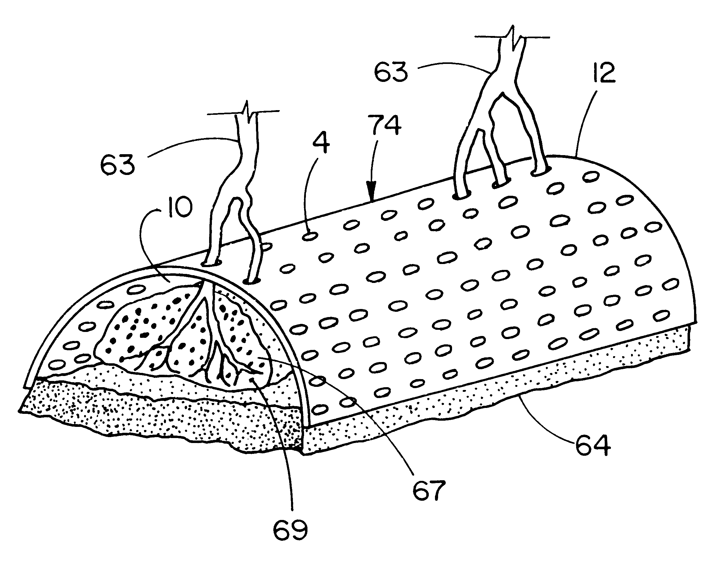
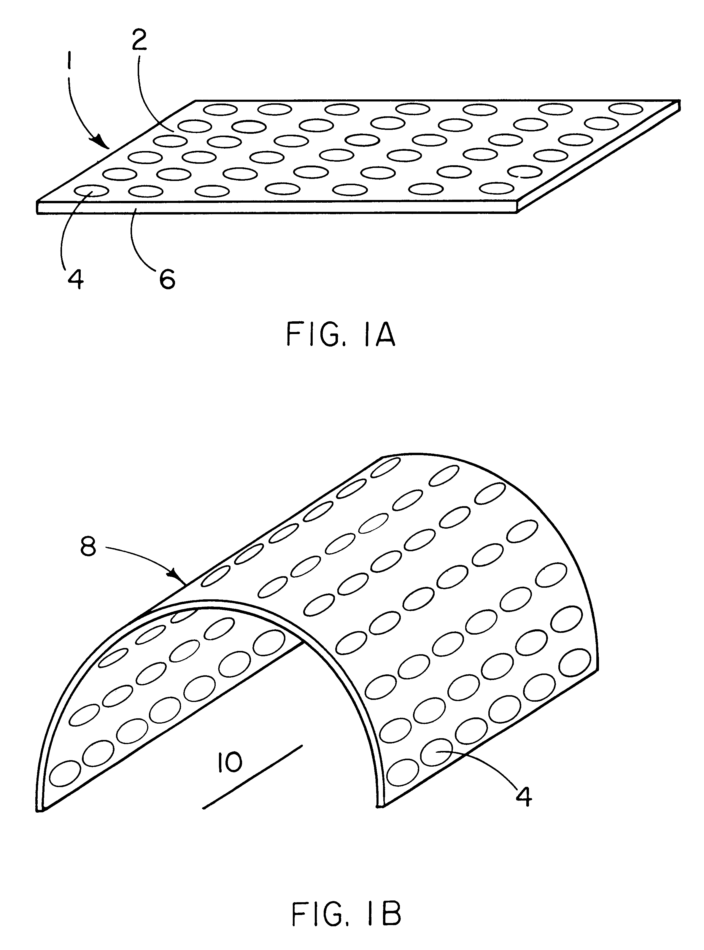
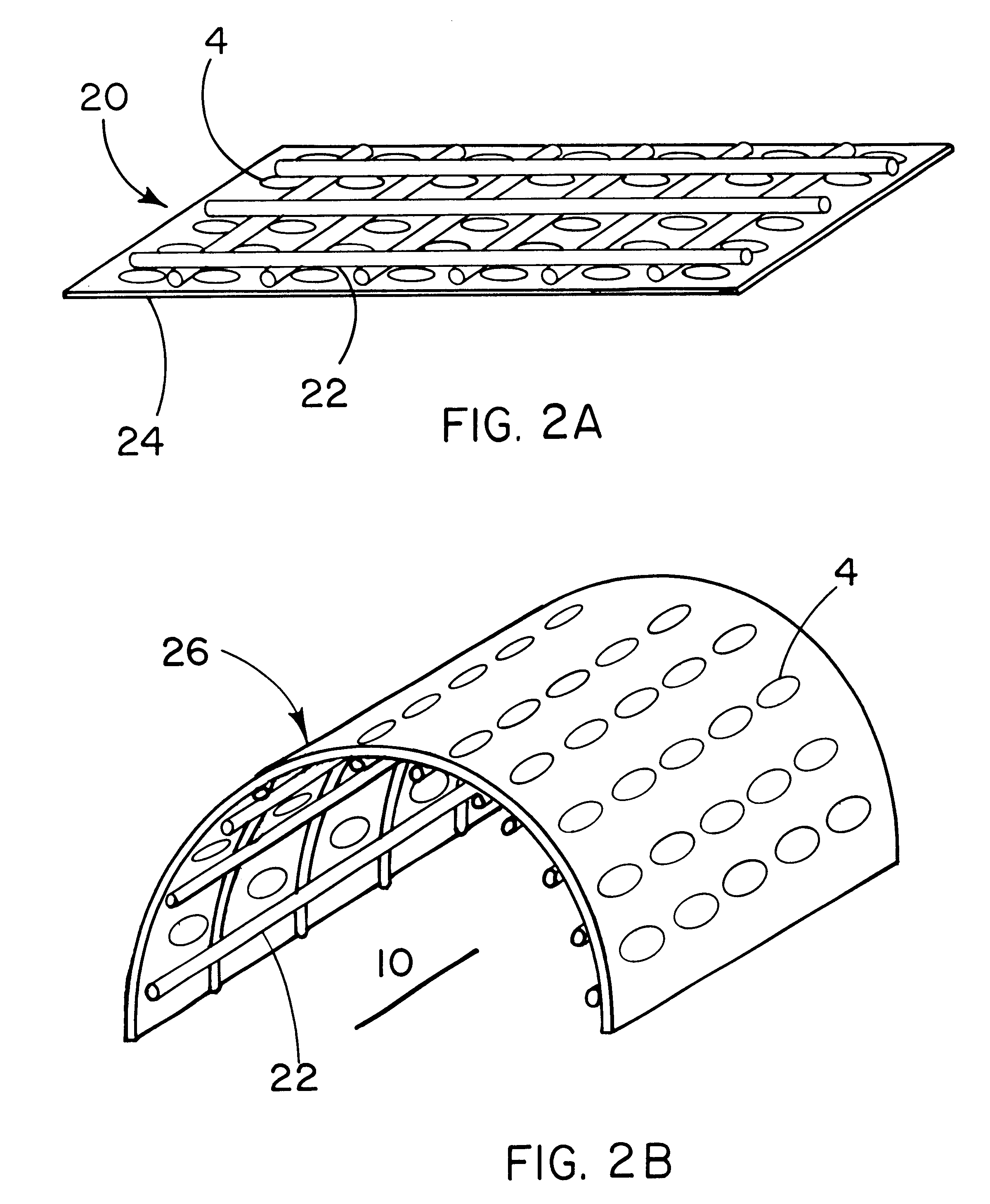
Current therapeutic options to increase bone or periodontal tissue mass are surgically complex and have several drawbacks.
The main drawbacks of therapies utilizing autograft bone are; the limited amount of tissue that is available for transfer, donor site morbidity and, in some cases, the complete lack of available or appropriate donor sites.
In addition, in the case of non-vascularized bone grafts, resorption of the transferred tissue can result in decreased tissue mass and inadequate function and / or aesthetic outcome.
Such procedures are not without problems however.
Associated problems with this technique may include lack of donors, immunological response of the host, revitalization of the grafted material by the host, and the possibility of disease transfer from donor tissue to the recipient.
However, these regenerative processes are dependent on, and limited by, both device design and the regenerative potential inherent in the biological processes of the individual.
This approach has been successful in instances associated with small defects but lacks the desired predictability of outcome in many clinically relevant large defects.
Interaction of the host cells, e.g. the so called foreign body reaction, with the porous matrix also may limit the rate, quantity and architecture of the bone formed within the device.
However, these materials also take up a significant portion of the space which could otherwise be occupied by newly formed bone.
As a consequence, such materials interfere with bone formation and also result in a potentially detrimental interface between bone and the biomaterial.
The presence of a biomaterial interposed within bone can interfere with normal bone remodeling processes and can ultimately result in bone resorption or stress fractures (Spector 1991).
Consequently, there is less space available for bone formation and significantly greater quantities of degradation products.
These degradation products can interfere with bone formation and can also result in bone resorption (Bostman 1992).
However, collagen sponges, especially those with suitable degradation time frames, are generally not able to withstand the above-listed in vivo stresses and consequently are unable to maintain the size and shape of the filled space.
As a result, the newly created bone assumes an ill-defined geometry which is not the same as the original shape of the sponge and which is often not of adequate or optimal functional or therapeutic benefit.
Wolfe and Cook (1994) have also recognized the difficulty of controlling the geometry of bone formed using osteogenic proteins and state that the osteoinductive effect of the protein can be difficult to confine to a limited anatomic area, especially using semi-solid carrier vehicles.
This same phenomenon can be seen with degradable synthetic polymer structures that, although they can be designed with appropriate mechanical stresses at the time of implantation, gradually lose their ability to withstand in vivo stresses and collapse at some point during degradation.
The desired bone tissue was therefore not formed and the cartilage tissue generated was associated with a non-degradable material that is likely to adversely influence the desirable properties of the natural tissue.
This technology has done much to advance the field of alveolar bone and periodontal tissue regenerative therapies, however, the widespread application of this technology has not been fully realized due to issues of predictability in the most clinically challenging situations.
In addition, studies by Hedner and Linde (1995) found that the combination of a cell and tissue excluding membrane and a bioactive molecule was less effective in stimulating bone healing in mandibular defects than the bioactive molecule alone.
One of the disadvantages of this technique is that it requires the use of living autogenous tissue, namely a muscle flap, which requires surgically injuring existing living tissue.
In addition, vascularized muscle tissue may not be available for transplantation in specific areas, such as the oral cavity, especially without significantly affecting other functions of the patient.
The mesh device used does not define the configuration of a space having the size and shape desired for the bone to be generated.
This study showed that the principles and mechanisms of guided bone regeneration do not operate when membranes are utilized with larger pore sizes that do not result in cell and tissue exclusion.
Consequently, significantly less bone regeneration results with large pore size membranes which do not exclude cells and tissue.
Although these studies showed that it is possible to regenerate bone in this manner, this approach also resulted in massive resorption of the graft.
It is clear that long bone defects do not heal appropriately in the long term using a combination of autograft bone and a single macroporous membrane.
An additional drawback of this methodology is that it requires the use of autologous tissue in quantities at least sufficient to fill the defect.
However, as the studies of Gugala and Golgolewski show, the use of a single macroporous sheet in conjunction with autograft bone does not result in a satisfactory long term healing response at least for long bone applications.
In addition, this approach also requires the use of autologous tissue to fill the defect.
It is technically difficult to achieve delivery of therapeutically effective quantities of an appropriate bioactive molecule from a membrane.
For example, it is not always possible to load sufficient quantities of a bioactive agent onto a membrane by relying on simple adsorption of the molecules onto the membrane surface, especially if the membrane is constructed from a hydrophobic material.
Even if sufficient loading of the membrane can be achieved, the next obstacle is providing an appropriate delivery profile.
It is not generally possible to achieve such a sustained release profile via simple adsorption and desorption of the bioactive molecule from a membrane, however.
In addition to the technical difficulties involved in achieving appropriate delivery of a bioactive agent from a membrane, it has not been shown that delivery of a bioactive molecule from a macroporous and tissue penetrable membrane surrounding the periphery of a bone defect is efficacious in regenerating bone.
First, the presence of the bioactive molecule in high concentrations near the outer surface of the membrane would be likely to cause bone to form on the outside of the membrane thus resulting in a lack of control of the regenerated bone geometry.
Second, diffusion of the bioactive molecule inward toward the center of the defect would result in an adverse concentration gradient for the migration of cells, such as mesenchymal stem cells, from the tissue surrounding the defect.
In each case, a bioactive agent delivered in an appropriate manner from a carrier material filling the space under a tissue excluding membrane did not achieve the desired bone regeneration result.
In addition, desired bone regeneration is not attained when a macroporous membrane, which does not result in cell and tissue exclusion, is used alone (Pineda et al.

Method used


Image

  • Methods and articles for regenerating bone or peridontal tissue
  • Methods and articles for regenerating bone or peridontal tissue
  • Methods and articles for regenerating bone or peridontal tissue

Examples


example 1

Periodontal Regeneration with Tissue Exclusive and Tissue Penetrable Devices

Purpose

The purpose of this study is to compare tissue-excluding (TE) and tissue-penetrable (TP) devices, for the ability to regenerate periodontal tissues in surgically created supraalveolar critical size defects. The devices are used to establish a space surrounding the teeth and surgically created defect. In this study, living tissue generation depends on the inherent biological regenerative potential of the subject. No exogenous TGF-Beta proteins or other bioactive molecules are placed in the space established by implantation of the devices in either experimental or control sites.

It is hypothesized that us a TP device, capable of allowing penetration of vascular structures and soft tissue cells into the established space, will result in regeneration of periodontal structures equivalent to TE devices.

Materials and Methods

Six male beagle dogs (age 18-24 months, weight approximately 15 kg), exhibiting intact...

example 2

Membrane vs Space Distribution Delivery of the TGF-Beta Protein rhBMP-2

Purpose

The objective of this experiment is to measure the release profiles of the TGF-Beta protein, rhBMP-2, from ePTFE and PGA:TMC membranes (membrane delivery). In addition, the membranes are treated with various agents to allow them to wet more easily, to change their surface chemistries, or to immobilize the protein through different types of bonds (e.g. ionic and covalent). The membrane release profiles resulting from these treatments are compared to those for a collagen sponge and a hyaluronic acid felt, which are chosen as appropriate carriers to deliver TGF-Beta proteins from a space established by a TP device (spatial delivery). The relative efficacy of spatial delivery versus membrane delivery is thus determined.

Materials and Methods

Material Description

Four materials were used as carriers in this release experiment: an expanded polytetrafluoroethylene (ePTFE) membrane, a membrane made from a poly(glycol...

example 3

Purpose

The objective of this study is to determine if the configuration (size and shape) of alveolar bone associated with critical size supraalveolar periodontal defects, generated in vivo under the influence of the TGF-Beta protein, recombinant human Bone Morphogenetic Protein-2 (rhBMP-2), can be predictably controlled with the use of a pre-configured TP, ePTFE device when the inductive protein is distributed throughout the space established by the device.

It is hypothesized that implantation of a TP ePTFE space-defining device will allow interaction of rhBMP-2, when delivered from within the space established by the device, with the target host cells and tissues in such a way that generation of alveolar bone tissue can be predictably controlled resulting in formation of bone tissue with desired configuration.

Materials and Methods

Four adult male or female mixed-breed dogs (age 10-12 months, weight $ 20 kg) are obtained from a USDA approved dealer. The animals exhibit intact mandibul...


PUM

PropertyMeasurementUnit
diametersaaaaaaaaaa
diametersaaaaaaaaaa
diametersaaaaaaaaaa

Abstract

There are numerous medical situations involving deficiencies of living bone or periodontal tissue and where increase of living bone or periodontal tissue mass is desired. Methods are described wherein a configured, shell-like device that is capable of being penetrated by living cells and tissues, is implanted into the body of a mammal in such a way as to establish a space, the space being at least partly, bounded by the device. The configuration of the device is such that the configuration of the established space is essentially the same as the configuration of living bone or periodontal tissue that is desired for treatment of the tissue deficiency. At least one protein from the Transforming Growth Factor-Beta Superfamily of proteins is placed within the established space for the purpose of stimulating the growth of living bone or periodontal tissue within the established space. A kit for the generation of living bone or periodontal tissue, comprised of the components mentioned above, is also disclosed.

Description

Current therapeutic options to increase bone or periodontal tissue mass are surgically complex and have several drawbacks. These options include the use of autografts, allografts, xenografts and alloplastic materials. Autografts involve the transfer of bone from one part of the patient to another (either as a vascularized graft or as a non-vascularized graft). The main drawbacks of therapies utilizing autograft bone are; the limited amount of tissue that is available for transfer, donor site morbidity and, in some cases, the complete lack of available or appropriate donor sites. In addition, in the case of non-vascularized bone grafts, resorption of the transferred tissue can result in decreased tissue mass and inadequate function and / or aesthetic outcome.When autograft bone is not available, allografts may often be used. Allografting involves the transfer of bone between two individuals of the same species. Such procedures are not without problems however. Associated problems with ...

Claims


Application Information

Patent Timeline
Patent Type & Authority Patents(United States)
IPC IPC(8): A61C8/00A61F2/28A61L27/38A61L27/48A61L27/00A61L27/54A61L27/56A61F2/00A61F2/02A61F2/30A61F2/46
CPCA61C8/0006A61F2/2846A61L27/3847A61L27/3865A61L27/48A61L27/54A61L27/56A61F2/2803Y10S623/901A61F2/468A61F2002/2817A61F2002/2835A61F2002/30062A61F2002/30153A61F2002/30235A61F2002/30238A61F2002/30785A61F2002/30787A61F2002/3082A61F2002/30909A61F2002/30914A61F2002/30948A61F2002/30971A61F2210/0004A61F2230/0019A61F2230/0069A61F2310/00365A61L2300/414A61L2300/64A61L2430/02A61F2002/30237
Inventor WHITE, CHARLES F.FLYNN, CHARLESCOOK, ALONZO D.HARDWICK, WILLIAM R.WIKESJO, ULF M. E.THOMSON, ROBERT C.
Owner WL GORE & ASSOC INC
Why Eureka
Try Eureka