
Rapid magnetic resonance imaging and magnetic resonance angiography of multiple anatomical territories

a magnetic resonance imaging and multiple anatomical technology, applied in the field of medical diagnostic procedures, can solve the problems of renal failure, inability to use intravenous contrast and image the blood vessels, and all these techniques have their own inherent limitations, and achieve the effect of low osmolality and high resolution

Inactive Publication Date: 2002-12-10
WILLIAM BEAUMONT HOSPITAL
View PDF18 Cites 49 Cited by
  • Summary
  • Abstract
  • Description
  • Claims
  • Application Information

AI Technical Summary

Benefits of technology

Therefore, it is desirable in the art of radiology to provide an imaging device which provides high resolution images and which permits rapid positional changes in patient centering so that the time required for obtaining accurate images of a patient may be reduced and so that movement of the patient relative to the imaging coils is simplified.
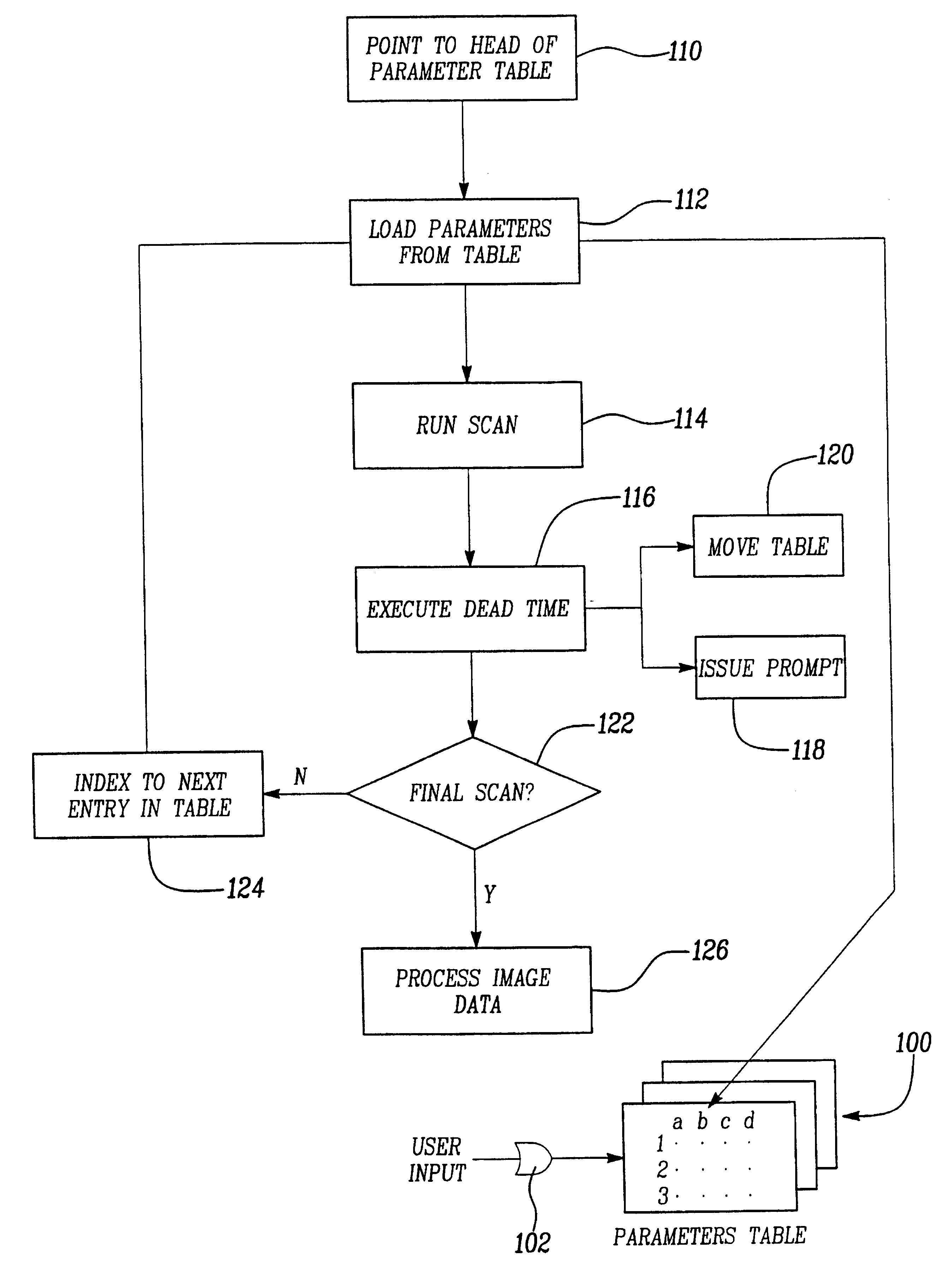
The present invention provides a procedure and apparatus which allows rapid positional change in the patient centering in order to facilitate the imaging of blood vessels in a series of different views, and to reduce the time required for obtaining the necessary images for a medical imaging examination using a single injection of an MRI contrast agent.
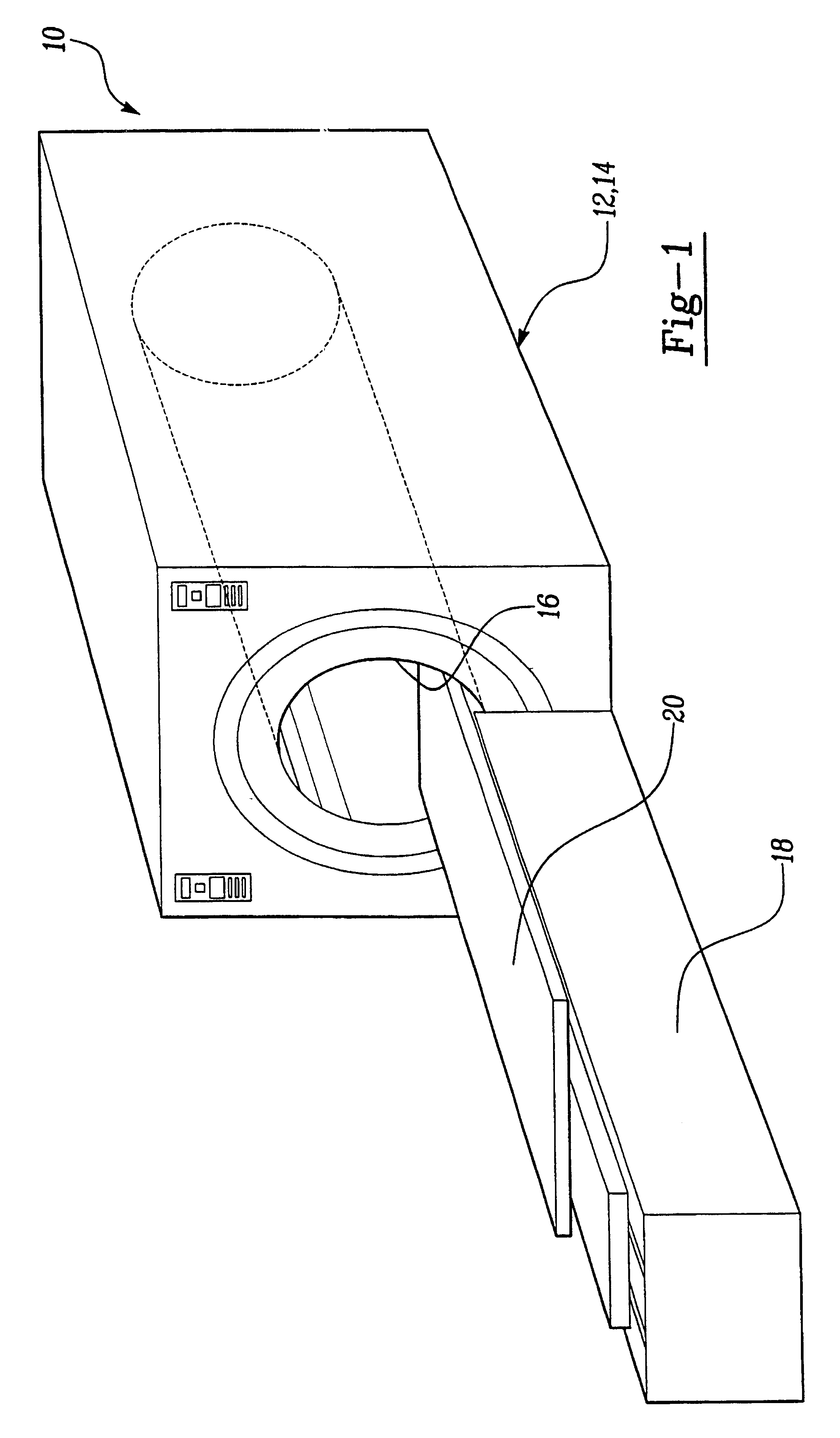
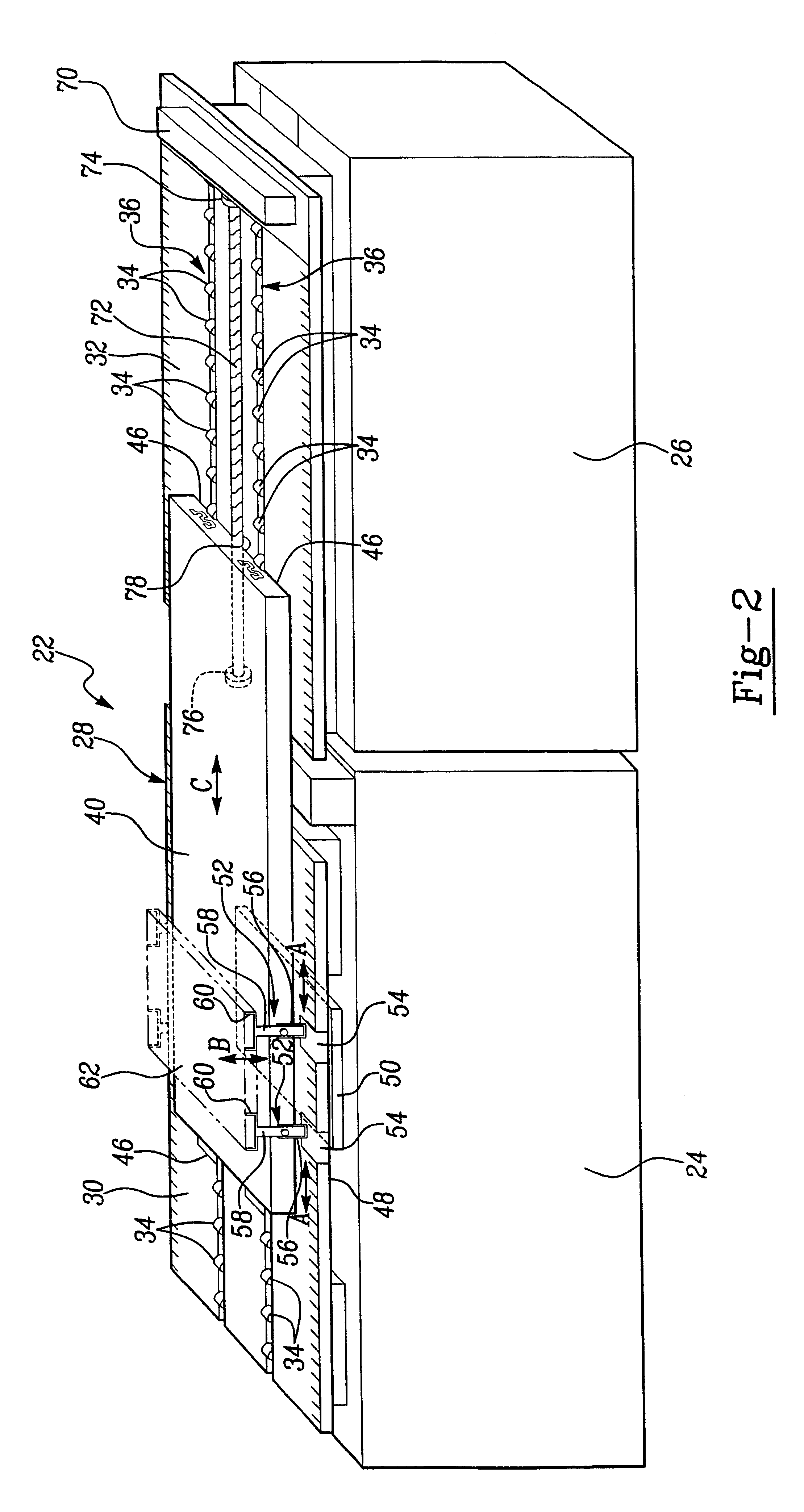
The kinematic imaging table of the present invention allows for the use of surface coils of an MRI machine for use in rapidly imaging different body locations in sequence including blood vessels and could therefore replace the predominant technique of x-ray angiography which is invasive, expensive and time consuming.
The dose of contrast and rate of injection of the contrast material varies among different contrast groups. Typically, the extracellular gadolinium chelates (Magnevist, Omniscan and ProHance) can provide adequate imaging of a target area with a dose of 0.1 mmol / kg and at a rate of 2-3 cc / sec.
Currently, the average hospital and professional fee for an x-ray aortogram with runoff evaluation is thirty five ($3500) hundred dollars. In comparison, the current average price for an MRI study employing one hour of table time is fifteen ($1500) hundred dollars. This price could also be lowered especially if imaging times are decreased with the current technology that is described herein. The cost should also decrease with newer contrast agents as described above. The blood pool agent, MS 325, can be administered at much lower doses. Likewise, the blood pool contrast agents have the potential in providing even higher signal-to-noise of the blood vessels, especially those that are much smaller in size as found in the most distal aspect of the lower extremities. When imaging different anatomical locations in sequence, however, one needs to alter various imaging parameters, specifically the center of data acquisition.
Finally, regarding contrast administration, a fixed rate and volume of contrast followed by a fixed rate and volume of saline flush can be delivered with the use of automated power injectors that are available on the market today for MRI and MRA studies. The use of power injectors enhance the reproducibility of studies and more accurately deliver the contrast required for imaging of blood vessels. Prior to contrast administration, a small test dose (2 cc) of contrast is typically administered and rapid imaging is performed at the level of interest over time (typically 30-90 seconds). The time of contrast arrival is determined from this test and is subsequently used to determine the onset of data acquisition following the intravenous administration of the remaining contrast dose (10-40 cc).

Problems solved by technology

When imaging multiple target sites, however, all these techniques have their own inherent limitations.
These invasive techniques have a number of complications that are related not only to arterial puncture but also to the iodinated contrast that is administered.
Iodinated contrast complications include allergic reactions ranging from minor skin reactions to anaphylactic shock and death, and renal failure from contrast induced nephrotoxicity.
These techniques do not use intravenous contrast and image the blood vessels due to the inherent contrast of moving blood on gradient echo sequences due to the flow related enhancement phenomenon.
Phase contrast techniques are likewise, time consuming.
As such, breath-hold contrast enhanced techniques have been published in the literature, however, these have only been used in studying one anatomic region.
The limitations of contrast-enhanced techniques for evaluating multiple anatomical locations, therefore, are primarily related to the current inability of any of the MRI machines available on the market in rapidly moving the patient table to image successive anatomical locations.
Existing MRI machines are provided with movable tables, however, these tables and their respective control systems are designed to lock the table in place during a scan sequence and do not permit the examination table to be moved without requiring the MRI device to be recalibrated after the examination table is moved.
Furthermore, the existing CT or MRI machines are not capable of rapid positioning for patient centering in order to allow the patient to be repositioned for each scan sequence.
Using conventional techniques, this requires a considerable amount of time including moving the surface coils that are placed anterior and posterior to the body to be properly aligned with the different anatomical location as well as the center of the magnetic field.
If there is motion of the patient in between the baseline and immediate post-contrast scans, there will be misregistration and inability to subtract the veins yielding suboptimal arterial phase images.
As such, this will lead to patient discomfort and potentially, non-diagnostic images.

Method used


Image

  • Rapid magnetic resonance imaging and magnetic resonance angiography of multiple anatomical territories
  • Rapid magnetic resonance imaging and magnetic resonance angiography of multiple anatomical territories
  • Rapid magnetic resonance imaging and magnetic resonance angiography of multiple anatomical territories

Examples


Embodiment Construction

Three dimensional magnetic resonance angiography (3D-MRA) techniques have been routinely used in the past for vascular imaging. In particular, 3D-MRA in a single breath-hold [Simonetti OP, Finn J P, White R D, Bis K G, Shetty A N, et al. ECG-triggered breath-held gadolinium-enhanced 3D MRA of the thoracic vasculature(abstr). In: Book of abstract: International society of Magnetic Resonance in Medicine 1996:703. Shetty A N, Shirkhoda A, Bis K G, Alcantara A. Contrast enhanced three dimensional MR angiography in a single breath hold: A novel technique. AJR 1995;165:1290-1292.] has been shown to be a very useful and rapid technique when used with paramagnetic contrast agents. Given the rapid nature of this new sequence, with the acquisition of an entire 3D-data set during the passage of MR contrast material through the arterial system, arterial phase imaging is possible. A delayed acquisition following the initial arterial phase results in the visualization of venous flow. Conventional...


PUM


Abstract

A procedure and apparatus are provided which allow rapid positional change in the patient centering in order to facilitate the imaging of blood vessels in a series of different views. This procedure and apparatus can also facilitate the imaging of other tissues of the body at different spatial locations as well. The procedure and apparatus reduce the time required for obtaining the necessary images for a medical imaging examination using a single injection of an MRI or iodinated contrast agent.

Description

1. Field of the InventionThe present invention relates generally to medical diagnostic procedures. More particularly the invention relates to use of a kinematic imaging table for rapid positional changes in patients' centering for use with a magnetic resonance imaging (MRI) machine. The kinematic imaging table is constructed of a non-magnetic material, so as not to interfere with the magnetic resonance imaging and may be motorized to allow automatic positional changes in patient centering under computer control during the diagnostic procedures. The diagnostic procedure entails the use of this table or other means to effect rapid positional changes, injection of an MRI contrast agent and the use of rapid imaging MRI sequences for the display of vascular territories along different anatomical positions of the body. According to another aspect of the present invention, a patient is injected with a contrast material, and multiple images are taken at different overlapping orientations us...

Claims


Application Information

Patent Timeline
IPC IPC(8): G01R33/54G01R33/563A61B5/055
CPCA61B5/0555A61B5/411G01R33/563G01R33/56375Y10S5/943A61B5/055A61B5/704
Inventor BIS, KOSTAKI G.SHETTY, ANIL N.
Owner WILLIAM BEAUMONT HOSPITAL
Learn More