DNA detector and DNA detection method

a detector and dna technology, applied in the direction of fluid pressure measurement, liquid/fluent solid measurement, peptide measurement, etc., can solve the problems of reduced detection sensitivity, high background level, and insufficient consideration of simultaneous processing of two or more samples, and achieve high-efficiency photodetecting

Inactive Publication Date: 2003-06-10
HITACHI LTD
View PDF19 Cites 3 Cited by
  • Summary
  • Abstract
  • Description
  • Claims
  • Application Information

AI Technical Summary

Benefits of technology

The first object of the present invention is to provide a solution to the above-said problems and to provide a method and small-sized device in which extra-sensitive DNA detection is made possible. To achieve the object, the present invention uses a He--Ne laser with an emission wavelength of 594 nm in DNA base sequencing determination by fluorescence detection type electrophoresis gel separation, and adopts a highly efficient photodetecting system.
Conversely, greater gap length will cause easier dispersion of the samples without migrating in a straight line in the gap; samples will not migrate in the others of the paired capillaries. The gap length of about 2 to 3 mm allows normal electrophoresis of the samples; however, normal electrophoresis may take place, depending on the conditions such as electrophoresis voltage. The gap length of about 1 mm is preferred in practice. That is, the gap length of 0.1 mm to 1.0 mm allows smooth and effective electrophoresis of the samples. Two or more optical detecting portions can be positioned in a straight line, and a single excitation light can be irradiated simultaneously on two or more optical detecting portions for fluorescence detection. Furthermore, two or more optical detecting portions are linked with each other through the solution, so the excitation light is not damped. This allows irradiation of the optical detecting portions with sufficient light intensity, providing high-precision highly sensitive fluorescence detection.

Problems solved by technology

The said examples of the prior art use one capillary, but sufficient consideration has not been given to simultaneous processing of two or more samples.
For fluorescence detection in the state of electrophoresis, generally, the background from the gel, scattered light from the inner and outer walls of the capillary as the gel support or fluorescence from the capillary itself is produced in addition to the fluorescence from the object fluorophore itself, resulting in higher background level and reduced detection sensitivity.
One of the major problems in ensuring highly sensitive fluorescence detection is how to cut off such backgrounds.
In practice, there remains a problem of how to cut off the background to achieve highly sensitive fluorescence detection, as mentioned above.

Method used


Image

  • DNA detector and DNA detection method
  • DNA detector and DNA detection method
  • DNA detector and DNA detection method

Examples


first embodiment

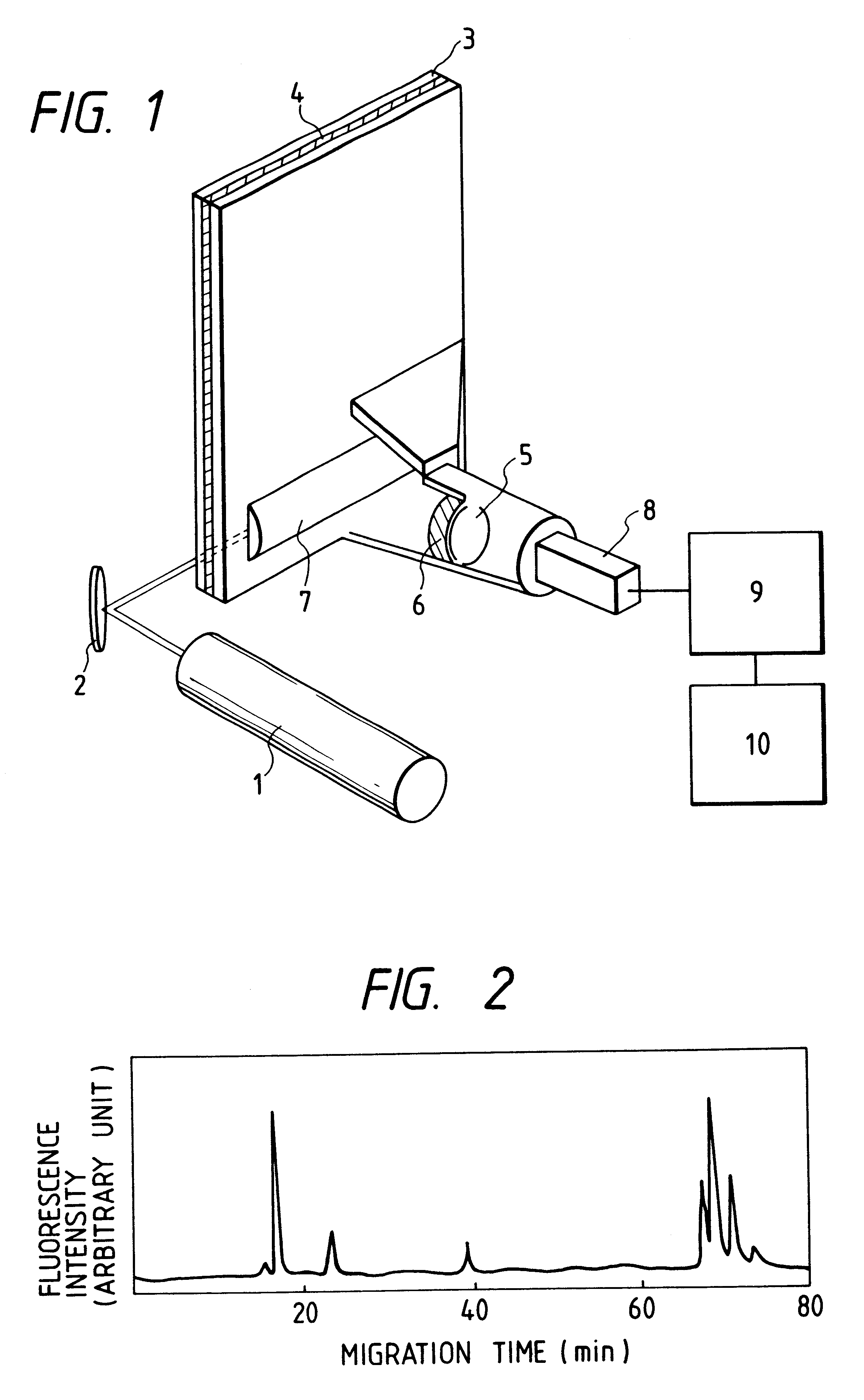
of the present invention will be described with reference to FIGS. 1 and 2. FIG. 1 is a schematic view representing the detector. Light emitted from the 594 nm He--Ne laser 1 irradiates the electrophoresis separation gel plate 4 from the side. After being collected by the cylindrical lens 7, the fluorescence emitted from the linearly irradiated portion is filtered out by the band pass filter 6, and forms images on the line sensor or secondary detector 8 through the lens for image formation 5. Filter 6 is a 6-cavity multilayer interference filter with a diameter of 50 mm, and transmittances for light having wavelengths of 594 nm, 600 nm, 610-630 nm and 640 nm are 10.sup.-4, 10.sup.-2, 0.6 or more, and 0.01 or less, respectively.

The concentration of the acrylamide gel constituting the gel plate 4 (concentration of the total quantity of monomer) is 4 to 6 percent (g / cc). When irradiated by the He--Ne laser with the wavelength of 594 nm, the background fluorescence from the gel has the ...

embodiment 2

In the present Embodiment, DNA fragments labeled by fluorescence are separated by electrophoresis and detection is made by fluorescence. The following describes the case of using Texas Red (Sulforhodamine 101, maximum emission wavelength of 615 nm) as labeling fluorophore. The DNA fragments as samples are labeled by fluophores. DNA fragments labeled by fluorophores are prepared by DNA polymerase reaction, using the primer labeled by fluorophore according to the well-known dideoxy sequencing method invented by Sanger and his colleagues. The details of the method of preparing samples according to Sanger's dideoxy sequencing method are described in the Embodiment 3.

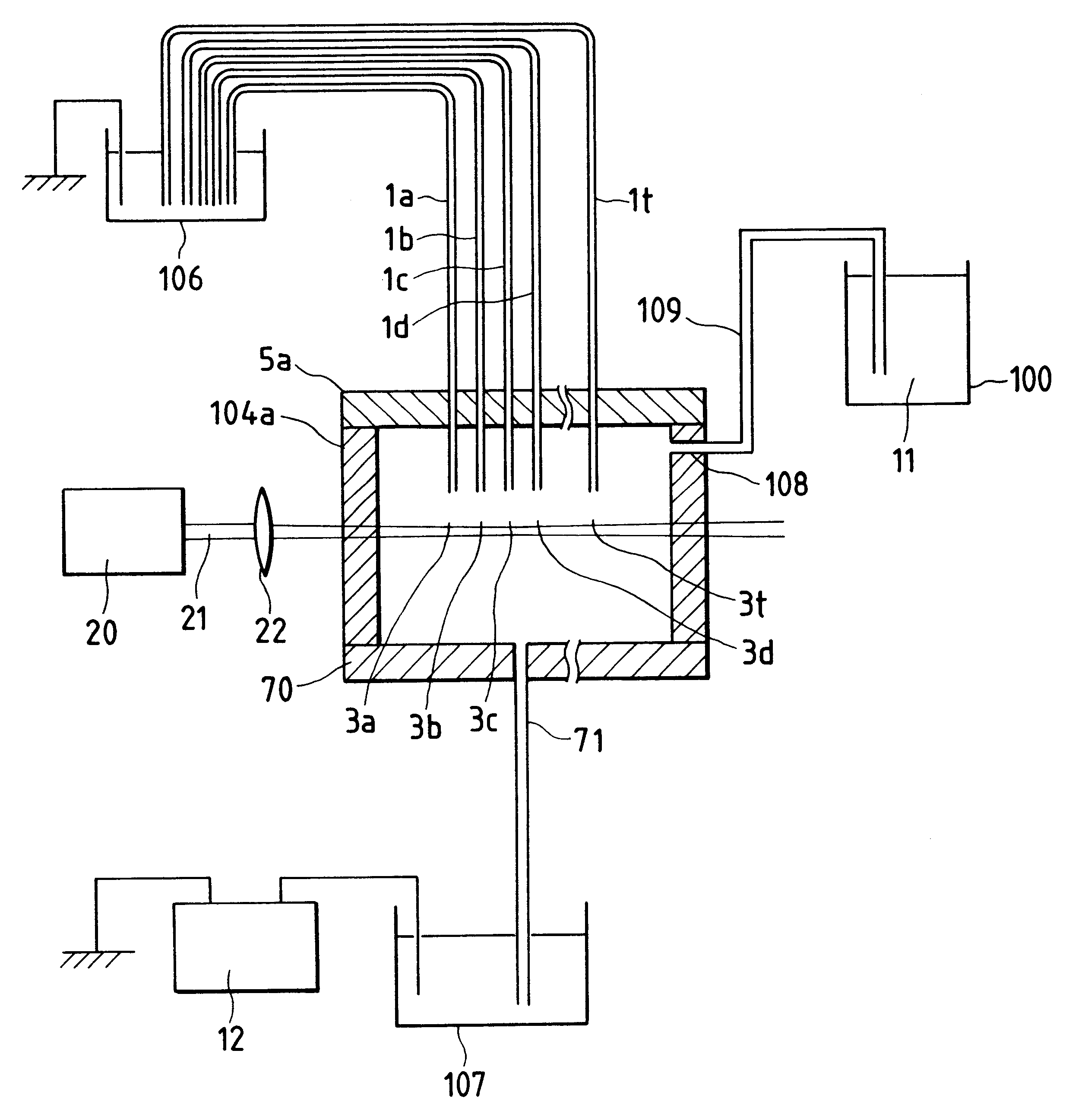
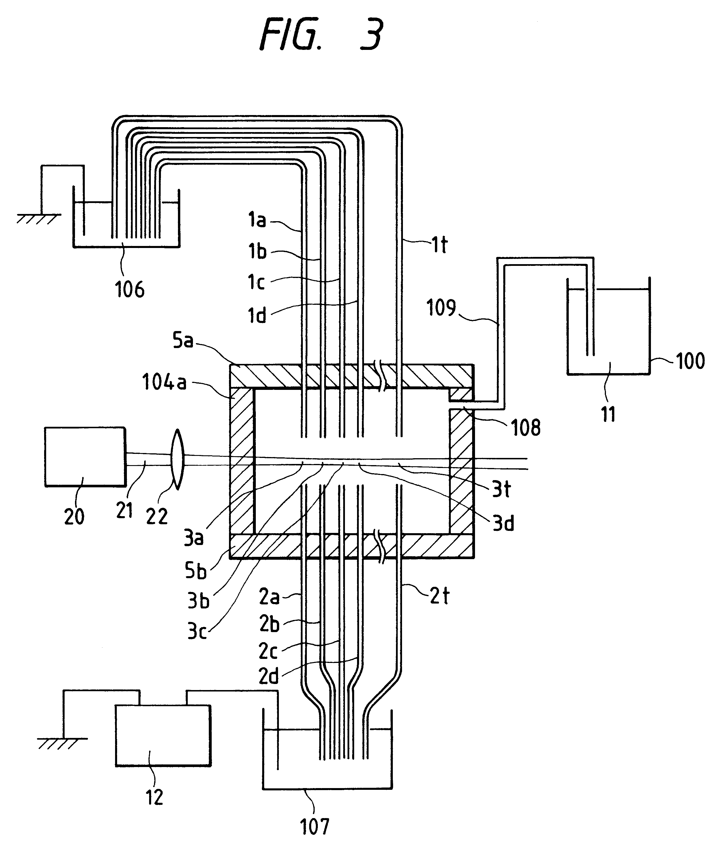
Firstly, the apparatus configuration will be described FIG. 3 shows the electrophoresis region and laser irradiation system of the electrophoresis apparatus according to the present Embodiment. FIG. 4 represents the fluorescence detection system of the electrophoresis apparatus. The electrophoresis apparatus includes an arra...

embodiment 3

The following describes the DNA sequence determination method using the apparatus introduced in Embodiment 2. DNA fragments labeled by fluorophores are prepared by DNA polymerase reaction, using the primer labeled by fluorophore according to the well-known dideoxy sequencing method invented by Sanger and his colleagues. The primer bonded with Texas Red (Sulforhodamine 101, maximum emission wavelength of 615 nm) is used as primer. Firstly, the labeled primer is added to the single-strand DNA and annealed, so that labeled primer is bonded to the single-strand DNA. This reaction solution is divided into four parts, which are subjected to DNA polymerase reactions corresponding to A, C, G and T, respectively. That is, four types of deoxynucleotide triphosphates (dATP, dTTP, dCTP and dGTP) and ddATP which will be a terminator are added to the single-strand DNA bonded with labeled primer to cause DNA polymerase reaction. The above procedure provides DNA fragments labeled by fluorophores ha...


PUM

PropertyMeasurementUnit
wavelengthaaaaaaaaaa

Abstract

An electrophoresis apparatus includes an optical cell having an outlet for fluid and filled with a buffer solution, and a plurality of electrophoresis lanes, each including a separation medium, which separate samples labeled with fluorophores, one end of each of the electrophoresis lanes being disposed in the optical cell to enable the separated samples labeled with the fluorophores to migrate from the electrophoresis lanes into the buffer solution in the optical cell. There is also provided a vessel containing a buffer solution, the vessel being disposed so that a level of the buffer solution in the vessel is higher than the ends of the electrophoresis lanes disposed in the optical cell, a passage connecting the vessel with the optical cell, a light source which emits light which excites the fluorophores to emit fluorescence, and a photodetector which detects the fluorescence emitted by the fluorophores.

Description

The present invention relates to a method of detection of DNA and protein and of DNA base sequencing determination and to an apparatus therefor.It relates more particularly to the fluorescence detection type gel electrophoresis apparatus.For DNA base sequencing determination by eletrophoresis gel separation, a radioisotope label has been used as a label for a DNA fragment. Due to the inconvenience of this method, however, a method of using a fluorescence label has come to be increasingly employed. (Refer to U.S. patent application Ser. No. 07 / 506,986 (U.S. Pat. No. 5,062,942) and Bio / Technology Vol 6, July 1988, pp816-821, for example.) As an excitation light source this method uses an argon laser with an output of 20 to 50 mw and a wavelength of 488 nm or 515 nm to detect the DNA fragment of 10.sup.-16 mole / band to 2.times.10.sup.-18 mole / band. As fluorophores, the method has used FITC (fluoresce in isothiocyanate with a maximum emission wavelength of 515 nm), SF (succinyl fluoresc...

Claims


Application Information

Patent Timeline
Patent Type & Authority Patents(United States)
IPC IPC(8): G01N27/447G01N35/00
CPCG01N27/44721G01N27/44782G01N2035/00237
Inventor TAKAHASHI, SATOSHIKAMBARA, HIDEKI
Owner HITACHI LTD
Why Eureka
Try Eureka