Assay devices and methods of analyte detection

analyte detection and assay device technology, applied in the direction of biomass after-treatment, analysis using chemical indicators, instruments, etc., can solve the problems of short time, practical limitations of the use of assay devices, and the sensitivity of such devices is often questioned, so as to improve the sensitivity of the assay, and improve the titration end-point activity

Inactive Publication Date: 2005-02-01
GENELABS DIAGNOSTICS PTE
View PDF34 Cites 70 Cited by
  • Summary
  • Abstract
  • Description
  • Claims
  • Application Information

AI Technical Summary

Benefits of technology

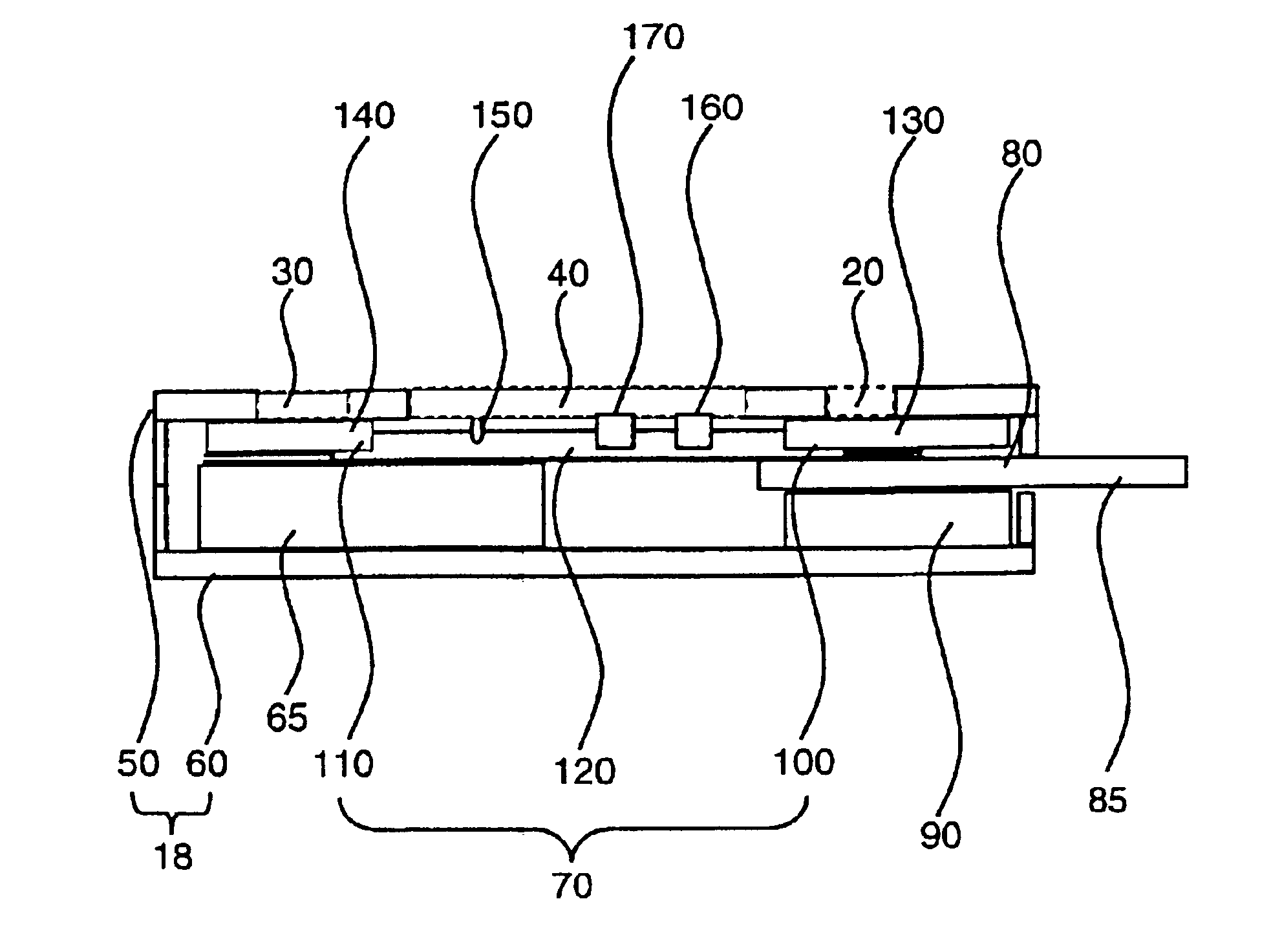
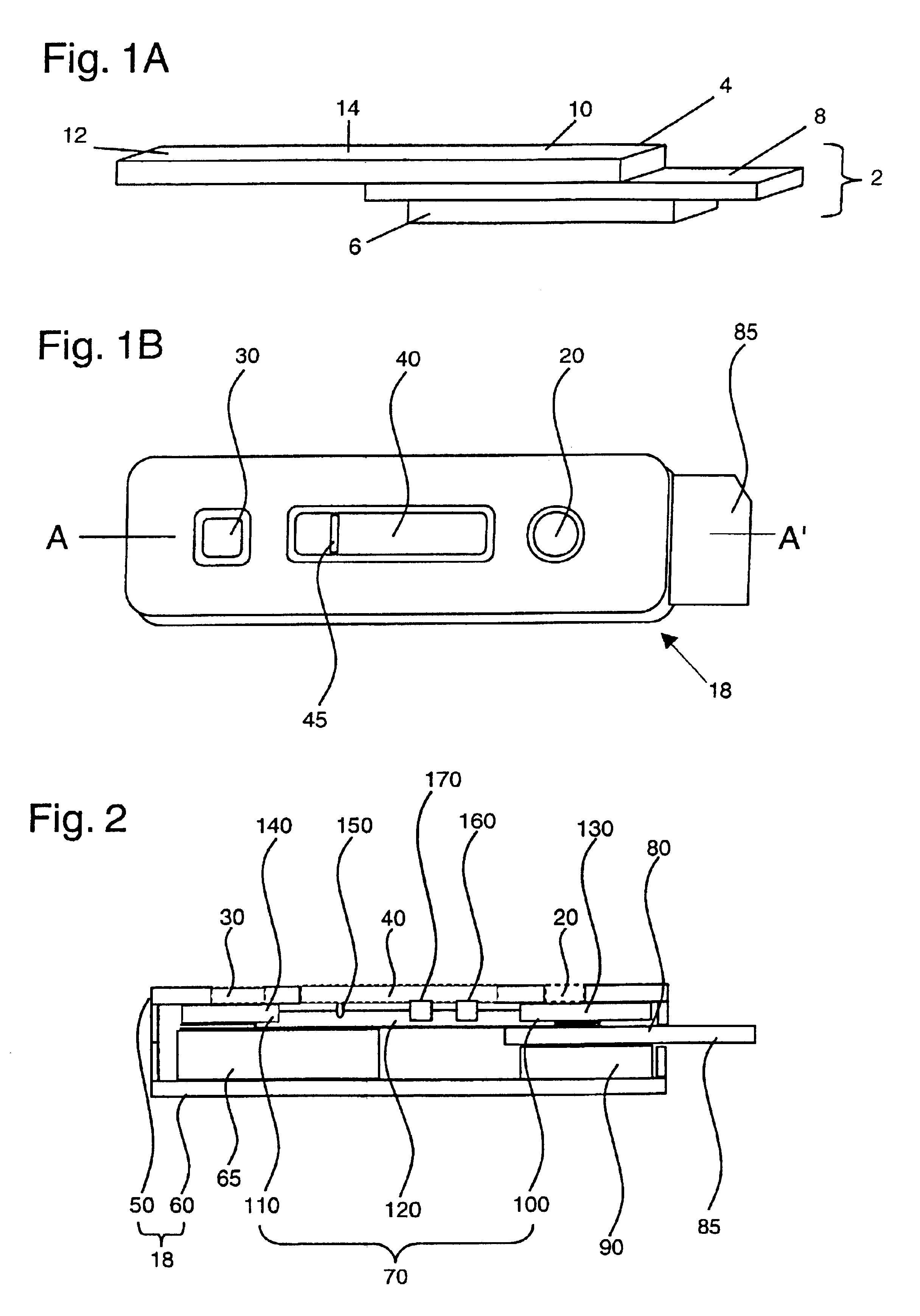
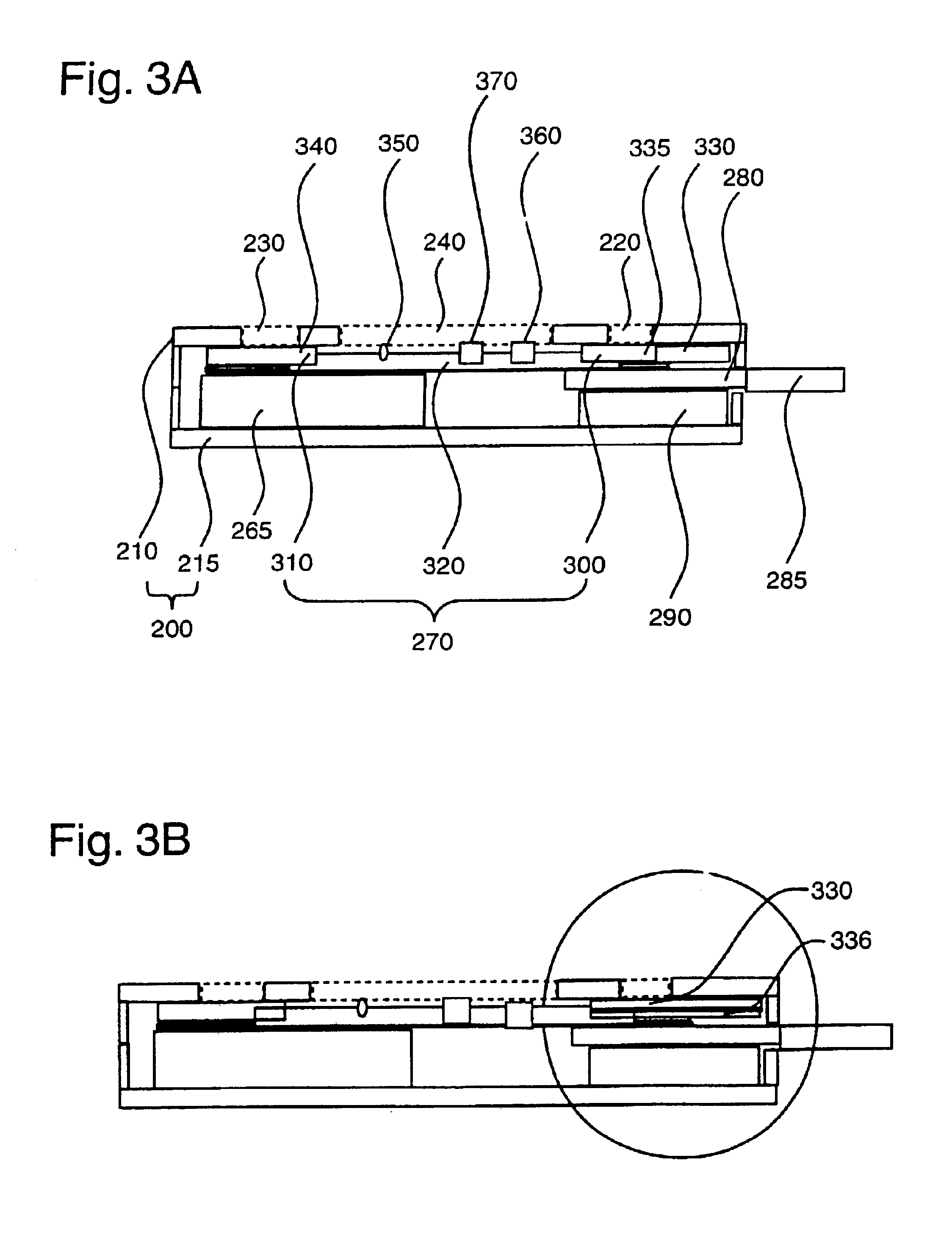
[0014]The present invention provides novel assay devices, test kits, and methods for detecting the presence of one or more analytes in a sample. The novel approach of the present invention provides optimal control of the assay reactions without requiring specially-developed specific antibodies, large volumes of sample, or complicated arrays of reagents or fluid pathways (for example, as compared to that described in U.S. Pat. Nos. 4,960,691 and 5,607,863). The present invention presents assay devices that are particularly suitable for rapid chromatographic assays using a controlled series of reactions. By controlling the release of the different reagents used in the assay device, the sensitivity of the assay is improved as compared to conventional assays, without compromising the specificity of the assay. The assay devices use a small volume of sample and achieve a much higher titration-end-point activity than conventional lateral flow assays. In addition, the assay devices of the present invention provide better assay sensitivity, without compromising specificity, a highly desirable improvement in the field of rapid chromatographic detection. In addition, the assay can be performed by untrained personnel in a minimum amount of time, and without the need for specialized equipment.

Problems solved by technology

The conventional rapid assays are a popular choice for determining the presence of a given analyte in samples provided at the “point of care” sites because they are relatively easy to use, do not use specialized equipment or personnel, and produce results in a short amount of time.
Most importantly, the sensitivity of such devices has often been questioned, due to various limitations with the currently available formats (Giles et al.
In addition, there are several practical limitations to the use of these assay devices inherent in the design of the assay format, as exemplified below.
This is a considerable limitation to use of such a device for analysis of serum or blood samples.
However, the flow-through format cannot be employed in a truly self-contained device.
This leads to additional limitations regarding reagent stability, if the detecting reagents are provided in liquid form, or issues surrounding the proper preparation and handling of the detecting reagent, if provided in a dried form.
However, the lateral-flow format does not allow for a washing step, as inherent in the flow-through format.
Any interfering species, such as particulate or colored material introduced by the sample solution, or unbound label, can potentially interfere with the readout of the assay device.
On the other hand, the near-patient whole blood or serum / plasma based rapid test devices that have recently become available have not lived up to expectations.
Although some of these rapid test kits perform with approximately 90% sensitivity, often these same kits are compromised by lower performances in specificities, especially when used in different geographical territories.
Furthermore, the sensitivities of standard serological tests are often heavily diminished in tuberculosis patients co-infected with HIV.

Method used


Image

  • Assay devices and methods of analyte detection
  • Assay devices and methods of analyte detection
  • Assay devices and methods of analyte detection

Examples


example 1

[0078]Assay devices for the detection of human antibodies to HIV types 1 and 2 were prepared as follows. Recombinant HIV 1 antigens p24 and gp41, and recombinant HIV 2 antigen gp36, were immobilized, or “slotted,” at a concentration range of about 0.08 to about 0.3 mg / ml onto a nitrocellulose membrane of 8 μm average pore size (Whatman, Ann Arbor, Mich.) using an IVEK (IVEK Corporation, N. Springfield, Vt.) Multispense 2000 striping machine. Protein A was immobilized in the same manner for use as an assay control line. The membrane was dried for approximately 10 minutes before addition of a blocking buffer (Milli-Q purified water with 0.3% casein and 0.25% sucrose). The membrane was exposed to the blocking buffer for approximately 1 minute, after which the membrane was dried at 37° C. for another 15 minutes. The membrane was finally affixed to a membrane backing (Adhesives Research Inc., Glen Rock, Pa.).

[0079]A reagent-bearing pad was prepared using a porous matrix (Hollingsworth & ...

example 2

[0083]Assay devices for the detection of human antibodies to Helicobacter pylori were prepared in a manner similar to that described in Example 1. Briefly, a nitrocellulose membrane of 81 μm average pore size was slotted with one or more native or recombinant antigens of H. pylori at a concentration of approximately 0.6 mg / ml using the IVEK striping machine. Exemplary recombinant antigens include, but are not limited to, the H. pylori antigens described in WO98 / 49314. The membrane was dried for 10 minutes before blocking with a blocking buffer (Milli-Q purified water with 0.3% casein and 0.25% sucrose) for 1 minute. The blocked membrane was then dried at 37° C. for another 15 minutes before being affixed to a membrane backing. A reagent-bearing pad was prepared using a porous matrix and sprayed with goat anti-human IgG antibodies that were labeled with colloidal gold particles of approximately 40 nm. The reagent-bearing pad was also dried at 37° C. for 30 minutes prior to use. The c...

example 3

[0087]An experiment was also performed with the H. pylori assay device as prepared in Example 2 using saliva samples instead of serum samples. Saliva samples from a healthy individual and an H. pylori infected individual, as confirmed by a Western blot test, were collected and diluted 1:5 in phosphate buffer saline (0.01M, pH 7.4). The samples were centrifuged for 5 minutes at 12,000 rpm and then stored in a freezer at −20° C. before use. Approximately 30 μl of each sample was applied to separate assay devices and tested according to the assay procedure described in Example 2. As a negative control, an assay using only phosphate buffer saline (0.01M, pH 7.4) was included in the experiment. The saliva sample from the infected individual produced a defined control band as well as an infection-indicating band with intensity in the range of 1+ to 2+. Neither the saliva sample from the healthy individual nor the PBS experiment control gave rise to an infection-indicating band. The saliva...


PUM

PropertyMeasurementUnit
volumesaaaaaaaaaa
volumesaaaaaaaaaa
volumesaaaaaaaaaa

Abstract

Assay devices, kits, and methods for detection of one or more analytes in a sample are provided. The assay device features the controlled release of reagents and hence is particularly suitable for binding assays such as immunoassays. The assay device achieves greater sensitivity than conventional rapid test assays, leading to stronger and/or more stable visual signals than those produced by conventional devices, easier interpretation of results, and reduced occurrence of indeterminate results. The device can be used for detecting analyte in a variety of biological samples without the need for conventional sample filtration techniques, and thus is suitable for use by untrained personnel without specialized equipment. In addition, the device can be used to simultaneously analyze a number of analytes using a single sample.

Description

CROSS-REFERENCES TO RELATED APPLICATIONS[0002]This application is a divisional continuation-in-part of and claims the benefit of U.S. application Ser. No. 09 / 771,479 filed Jan. 25, 2000, now U.S. Pat. No. 6,617,116, which is a continuation in part of U.S. Ser. No. 09 / 493,408 filed Jan. 28, 2000, now U.S. Pat. No. 6,316,205, the disclosures of which are incorporated by reference for all purposes.BACKGROUND OF THE INVENTION[0003]Chromatographic assay systems employed as rapid assay devices are one of several means for detecting the presence of a given analyte in a biological sample. One advantage to these systems is that the execution of these assays does not use additional specialized equipment or trained personnel. Another advantage is the great variety of analytes that can be detected using this type of assay. The use of rapid chromatographic techniques for detection of the presence of an analyte in a biological sample has thus progressed beyond the bounds of the clinical laborator...

Claims


Application Information

Patent Timeline
Patent Type & Authority Patents(United States)
IPC IPC(8): G01N33/558
CPCG01N33/558G01N2333/16G01N2333/205Y10S436/81Y10S435/81Y10S435/97G01N2333/35G01N33/54388
Inventor GUAN, MINGCHEN, HSIAO YINGCHOW, THERESA PUIFUNPEREIRA, ADRIAN RENNIEMUN, PING KUEN
Owner GENELABS DIAGNOSTICS PTE
Why Eureka
Try Eureka