Device for monitoring cells

a cell and cell technology, applied in the field of cell monitoring devices, can solve the problems of requiring a long incubation period, affecting the detection, identification and analysis of the behavior of microorganisms, and many procedures still time-consuming and laborious

Inactive Publication Date: 2005-05-31
BECTON DICKINSON & CO
View PDF8 Cites 51 Cited by
  • Summary
  • Abstract
  • Description
  • Claims
  • Application Information

AI Technical Summary

Benefits of technology

[0025]It is therefore an object of this invention to provide an improved means to detect the presence of, and to evaluate the metabolic activity of, eukaryotic cells present in a liquid or semi-solid media. It is further an object of this invention to provide a microbial monitoring device or system which can be simply read and visually interpreted, and which permits results to be obtained in a shorter time period than previously attainable, nominally 6 hours or less. Additionally, it is an object of the invention to provide a means for detection and / or monitoring the activity of eukaryotic cells without the use of dedicated instrumentation.

Problems solved by technology

While the science of microbiology has changed significantly in the last 25 years, many procedures for the detection, identification and analysis of the behavior of microorganisms are still time consuming.
However, this test method suffers from the drawback of also requiring a long incubation period.
However, such a system is “destructive” in nature, requiring the spraying of a developing solution of a color forming dye onto the test plate.
Re-incubation and reading at a later time is, thus, not possible and if the rapid technique fails, the experiment cannot be continued for a standard evaluation at a later time.
However, the procedure tends to be cumbersome and broad applicability has not been shown.
This method measured the change in oxygen concentration in a slow and cumbersome manner.
Unfortunately, the Clark electrode consumes oxygen during use (thereby reducing the oxygen available to the microorganisms) and the “standard” size electrode is typically used only to measure volumes of 100 mls or greater to prevent the electrode from interfering with the measurements.
Optical techniques allowing both colorimetric and fluorometric analyses for oxygen to be carried out rapidly and reproducibly are known, and costs for such analyses are often quite low.
However, such methods have not been adapted to microbial monitoring or prokaryotic or eukaryotic cell monitoring.
The increased incidence of tuberculosis and the recent emergence of Multiple Drug Resistant (MDR) strains threatens the ability to control this disease.
Many of these methods can only yield useful data when monitored by instruments dedicated to this task.
Among disadvantages of this method are the multiple reagent additions which are required.
MTT is also susceptible to interferences from some drugs with reducing groups and from precipitation of some drugs, especially those adsorbing light in the visible region.
The test itself is non-reversible and further time point readings of the same cell cultures cannot be performed without setting up a separate assay to be used for each time point.
Resazurin is subject to autoreduction in some media which can cause false positive signals.
This formulation, however, still requires the addition of dye and buffer to the cells and is essentially a non-reversible reduction.
This test is very sensitive but it is relatively expensive, time-consuming, and requires multiple steps.
This type of assay cannot readily be automated or adapted to formats for rapid drug screening purposes.

Method used


Image

  • Device for monitoring cells
  • Device for monitoring cells
  • Device for monitoring cells

Examples


example 1

Preparation of an O2-Sensitive Indicator Microtitration Tray

[0081]The fluorescent compound tris 4,7-diphenyl-1,10-phenanthroline ruthenium (II) chloride (Ru(DPP)3Cl2) was synthesized using the procedure of Wafts and Crosby (J. Am. Chem. Soc. 93, 3184(1971)). A total of 3.6 mg of the compound was dissolved in 2.0 ml dimethyl sulfoxide (D-5879, Sigma Chemical, St. Louis, Mo.) and the resultant solution was then added slowly, with stirring, to 1300 ml silicone rubber forming solution (Water Based Emulsion #3-5024, Dow Corning, Midland, Mich.). A 35 microliter aliquot of the mixture was subsequently dispensed into each well of a 96 well, flat bottom, white microtiter tray (#011-010-7901, Dynatech, Chantilly, Va.), and the system was subsequently cured overnight in a low humidity (less than 25% RH), 60° C. incubator. After curing, the trays were washed by either soaking or by filling and emptying each well several times with each of the following reagents; a) absolute ethanol, b)0.1 M ph...

example 2

Use of Indicator System to Measure Relative O2 Concentration Produced by a Reducing Agent

[0086]The O2 concentration in wells of an Indicator Microtiter tray produced as in Example 1 was varied by the addition of a strong reducing agent, sodium sulfite (which reduces O2 content). A 150 microliter aliquot of the reducing agent (at concentrations ranging from 0 to 1083 parts per million (ppm) sulfite ion in water) was pipetted into wells of the tray. Each well was allowed to react for 30 minutes, open to the atmosphere, and the fluorescence of the indicators measured in a Fluoroskan II Fluorometer (Flow Laboratories, McLean, Va.), having an excitation bandpass filter at a wavelength of 460 nm and an emission cut-on filter at 570 nm. The results are presented in Table 2.

[0087]

TABLE 2Effect of Sodium Sulfite on Fluorescenceppm sulfite ionFluorescence Intensity*030906535131633545325403354211571108311863*Mean of 4 wells

[0088]As shown, the wells containing the highest concentrations of red...

example 3

Use of Indicator System to Determine the Presence of a Microorganism

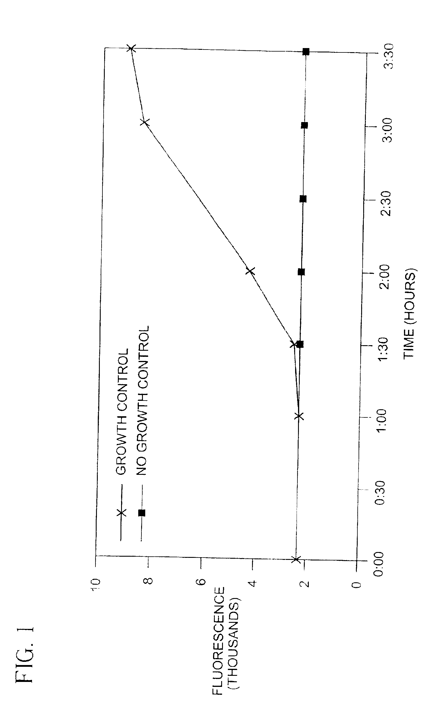
[0089]A 0.5 McFarland suspension of E. coli (ATCC #25922), containing about 1.5×10⁸ CFU / ml, was prepared using an A-Just nephelometer (Abbott Labs, Chicago, Ill.). The suspension was diluted to about 1×10⁷ CFU / ml in Broth A (see Example 1). A 150 microliter aliquot of this suspension was placed into indicator tray wells prepared as in Example 1, and subsequently incubated at 37° C. At intervals, the fluorescence was measured in a Fluoroskan II fluorometer over the period of 1-3½ hours. An increased fluorescence signal was observed over time as shown in FIG. 1. The fluorescence signal from wells containing no organisms showed very little change. The wells containing organisms were significantly brighter when visually observed under a UV light source. Thus, it appears that the metabolic activity of the organisms in the wells caused the fluorescence signal to increase (presumably by decreasing the O2 concentration).


PUM

PropertyMeasurementUnit
elapsed timeaaaaaaaaaa
time periodaaaaaaaaaa

Abstract

The present invention relates to methods for detection and evaluation of metabolic activity of eukaryotic and / or prokaryotic cells based upon their ability to consume dissolved oxygen. The methods utilize a luminescence detection system which makes use of the sensitivity of the luminescent emission of certain compounds to the presence of oxygen, which quenches (diminishes) the compound's luminescent emission in a concentration dependent manner. Respiring eukaryotic and / or prokaryotic cells will affect the oxygen concentration of a liquid medium in which they are immersed. Thus, this invention provides a convenient system to gather information on the presence, identification, quantification and cytotoxic activity of eukaryotic and / or prokaryotic cells by determining their effect on the oxygen concentration of the media in which they are present.

Description

[0001]This is a divisional of U.S. application Ser. No. 09 / 342,720, filed Jun. 29, 1999, which issued as U.S. Pat. No. 6,395,506 on May 28, 2002 and is a continuation-in-part of U.S. Ser. No. 08 / 715,557, filed on Sep. 18, 1996 now abandoned, which is a continuation-in-part of U.S. Ser. No. 08 / 025,899, filed on Mar. 3, 1993, which issued as U.S. Pat. No. 5,567,598 on Oct. 22, 1996, and which is a continuation of U.S. Ser. No. 07 / 687,359, filed on Apr. 18, 1991 now abandoned.BACKGROUND OF THE INVENTION[0002]1. Field of the Invention[0003]2. Description of Related Art[0004]Our environment contains a multitude of microorganisms with which we are continuously interacting. These interactions can be beneficial, i.e., fermentations to produce wine, vinegar or antibiotics; neutral; or even harmful, as in the case of infectious diseases. The ubiquitous presence of these microorganisms, thus, creates a continuing need for the detection, identification and study of the presence and metabolic ac...

Claims


Application Information

Patent Timeline
Patent Type & Authority Patents(United States)
IPC IPC(8): C12Q1/18C12Q1/04
CPCC12Q1/04C12Q1/18C12Q2304/44Y02A90/10
Inventor PITNER, J. BRUCEHEMPERLY, JOHN JACOBGUARINO, RICHARD D.WODNICKA, MAGDALENASTITT, DAVID T.BURRELL, GREGORY J.FOLEY, JR., TIMOTHY G.BEATY, PATRICK SHAWN
Owner BECTON DICKINSON & CO
Why Eureka
Try Eureka