

Determination of the ultrastructure of connective tissue by an infrared fiber-optic spectroscopic probe
a fiber-optic spectroscopic and ultrastructure technology, applied in the direction of optical radiation measurement, instruments, applications, etc., can solve the problems of difficult repair of articular cartilage defects, and achieve the effect of convenient evaluation and better treatment and managemen
- Summary
- Abstract
- Description
- Claims
- Application Information
AI Technical Summary
Benefits of technology
Problems solved by technology
Method used
Image

Examples
example 1
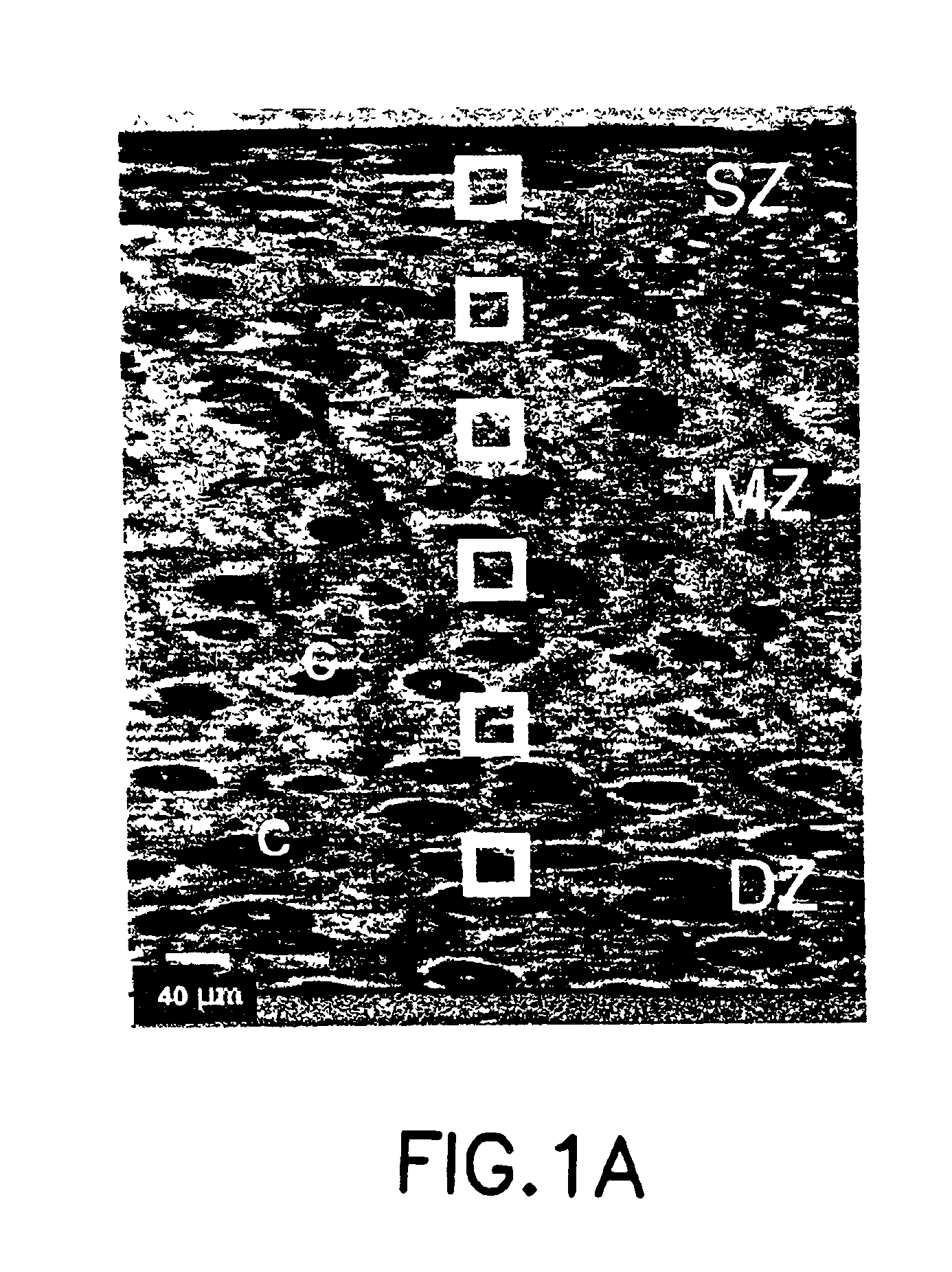
Cartilage Tissues
[0069]Full-depth cartilage explants were harvested from adult bovine occipital joints (obtained from local slaughter house) immediately after death. Six explants from two adult animals were examined. The tissues were removed from subchondral bone with a scalpel, and circular plugs (typically 0.5 to 1 mm thick) cut with an 8 mm diameter biopsy punch. The plugs were then snap-frozen in liquid nitrogen and stored at −70° C. For FT-IRM and FT-IRI analysis, the cartilage plugs were infiltrated with OCT embedding media (Miles Inc., Elkhart, Ind.) to facilitate sectioning, crysectioned at 6 μm thickness perpendicular to the articular surface, transferred to a BaF2 window and air-dried. Residual OCT in the tissue was dissolved with a few drops of water and the section re-dried.
example 2
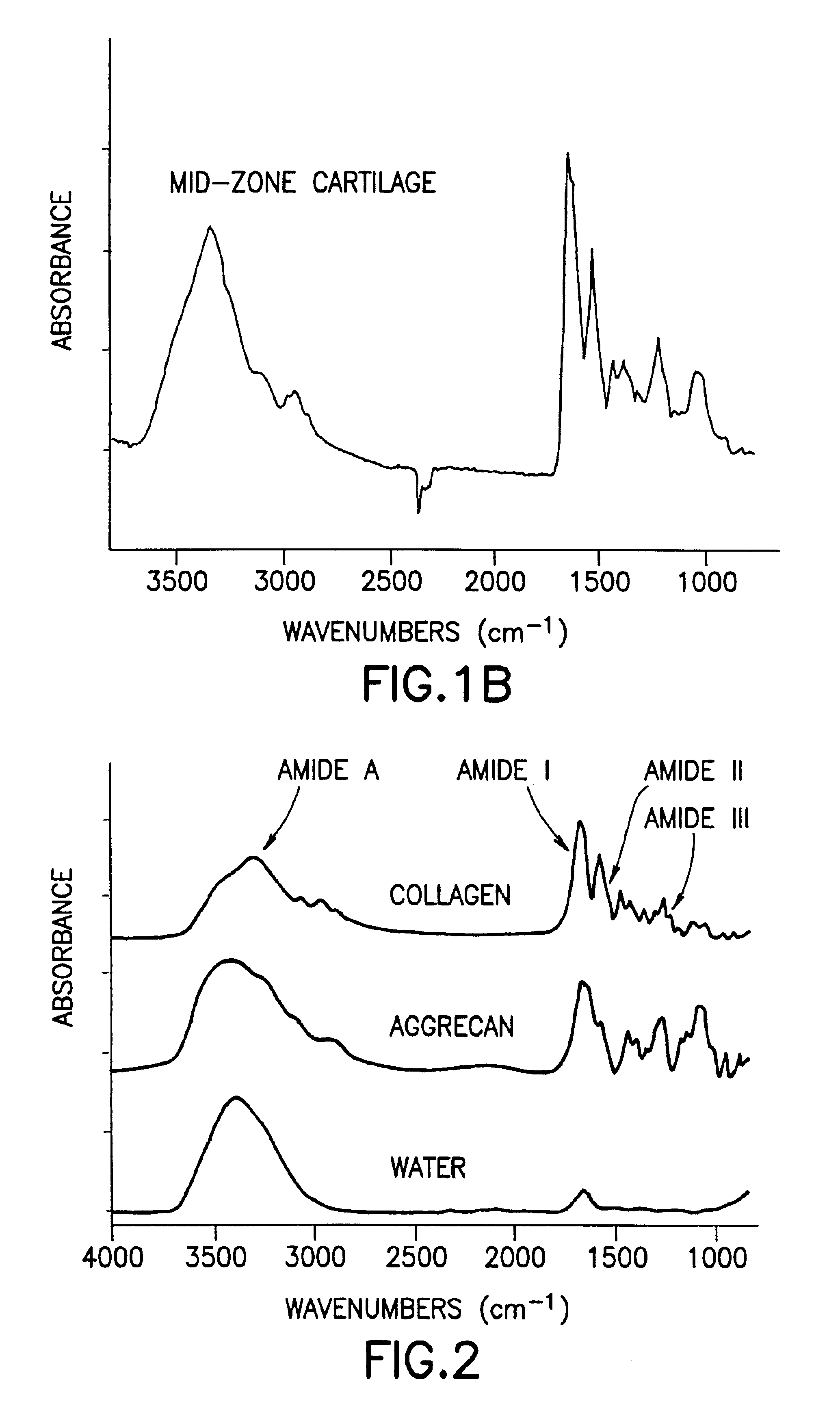
Model Compounds
[0070]Aliquots of chick type II collagen (Genzyme, Boston, Mass.) and purified calf nasal aggrecan were analyzed by FTIR and KBr pellets (2 mg sample: 200 mg KBr) using a Mattson Cygnus 25 Infrared Spectrometer (Mattson Instruments, Madison, Wis.). Ten sets of KBr pellets containing mixtures of type II collagen and aggrecan in varying proportions were also analyzed. The spectrum of liquid water was obtained by placing a drop of distilled water between two barium fluoride windows. Absorbance spectra were obtained by the co-addition of 256 interferograms collected at 4 cm resolution, followed by the Fourier transform of the resultant interferrogram.
example 3
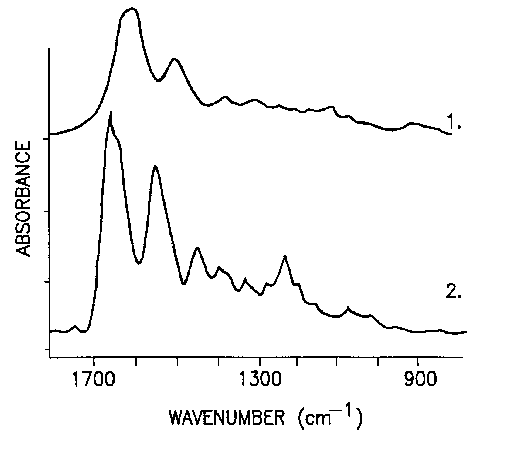
FT-IRM Analyses
[0071]A Bio-Rad (Cambridge, Mass.) FTS-40 infrared spectrometer coupled to a Bio-Rad UMA 500 microscope equipped with a mercury-cadmium-telluride (“MCT”) detector was used to acquire data at 4 cm−1 resolution under N2 purge. To obtain information on collagen and proteoglycan content and distribution, spectra of regions ranging from 20×20 μm to 40×40 μm diameter were acquired from all zones of cartilage. Typically, approximately 5 spectra per region of tissue analyzed were acquired.
[0072]To obtain information on the orientation of the collagen fibrils, polarization data was collected. A wire grid infrared polarizer was placed between the BaF2 window containing the tissue section and the impinging IR radiation. Spectral data was collected with infrared radiation polarized parallel and perpendicular to the cartilage articular surface. All data analyses were performed using Grams / 32 software (Galactic Industries, Salem, N.H.).
PUM
| Property | Measurement | Unit |
|---|---|---|
| diameter | aaaaa | aaaaa |
| length | aaaaa | aaaaa |
| diameter | aaaaa | aaaaa |
Abstract
Description
Claims
Application Information