Cells for detection of influenza and parainfluenza viruses
a technology for parainfluenza viruses and cells, applied in viruses/bacteriophages, instruments, recovery/purification, etc., can solve the problems of not being able to produce high-titer virus stocks for influenza, mink lung cells are expected to be less sensitive than desirable, and the virus produced from mink lung cells is not very infectious, etc., to achieve the effect of increasing the sensitivity of at least and enhancing the productivity of infectious viruses
- Summary
- Abstract
- Description
- Claims
- Application Information
AI Technical Summary
Benefits of technology
Problems solved by technology
Method used
Image
Examples
example 1
Generation of Transgenic Mink Lung (Mv1Lu) Cells Which Express Human Furin (hF)
[0084]Monolayers of mink lung cells (ATCC # CCL-64) were subcultured after treatment with trypsin, seeded into wells of 12-well plates in the presence of E-MEM culture medium (Diagnostic Hybrids), and incubated at 36° C. incubator for 48 hr. The freshly formed cell monolayer was subsequently used for transfection.
[0085]The human furin gene (GenBank Accession Number X17094; and FIG. 4, Panel A) was cloned in pcDNA3 using standard molecular biological techniques. The pcDNA3 plasmid which is commercially available from Invitrogen, is a standard eukaryotic expression vector with a cytomegalovirus enhancer / promoter flanking the multiple cloning site, and containing a neomycin-resistance cassette permitting the selection for stable transfectants. The plasmid DNA of pcDNA3-hF obtained from Dr. Nabil G. Seidah (Clinical Research Institute) was used to transform E. coli, and bacterial colonies which contained the ...
example 2
Detection of Influenza A Virus From Clinical Samples Using Mv1Lu-hF Cells
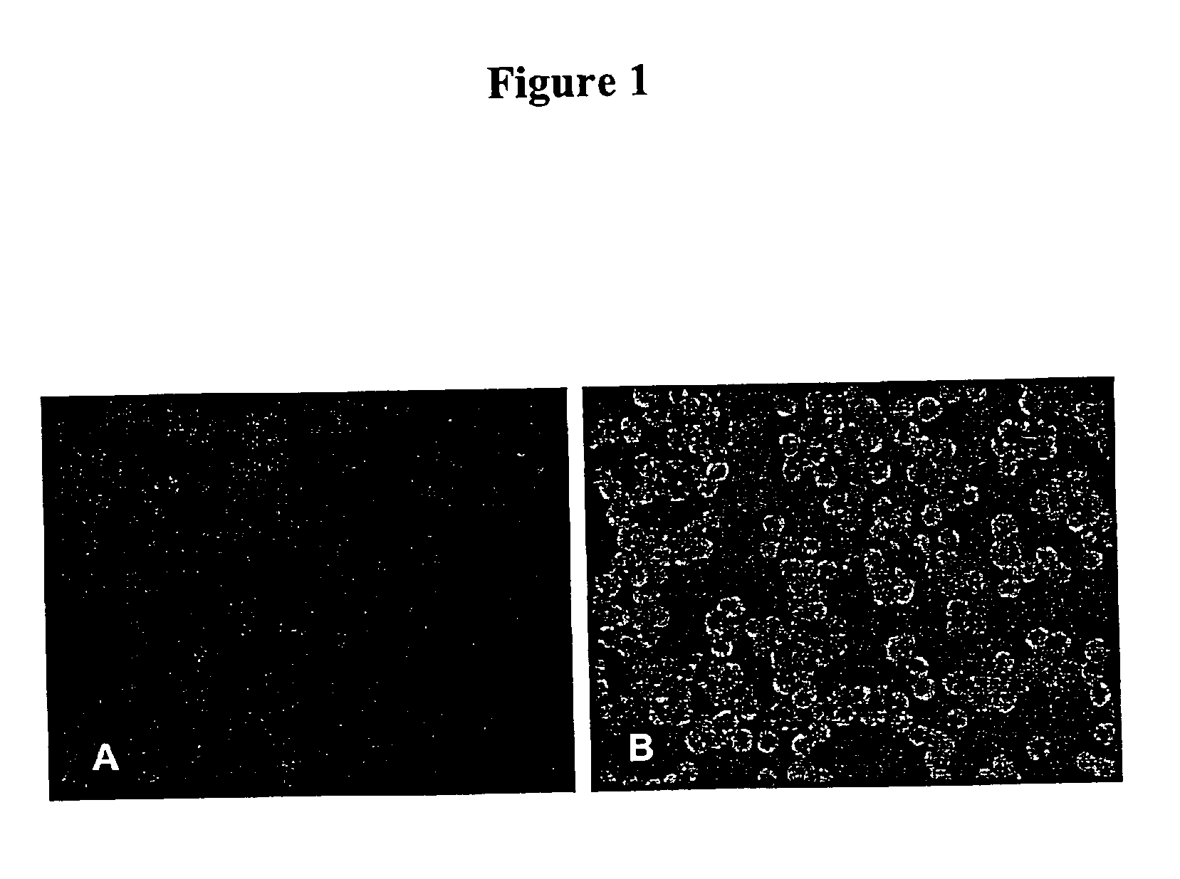
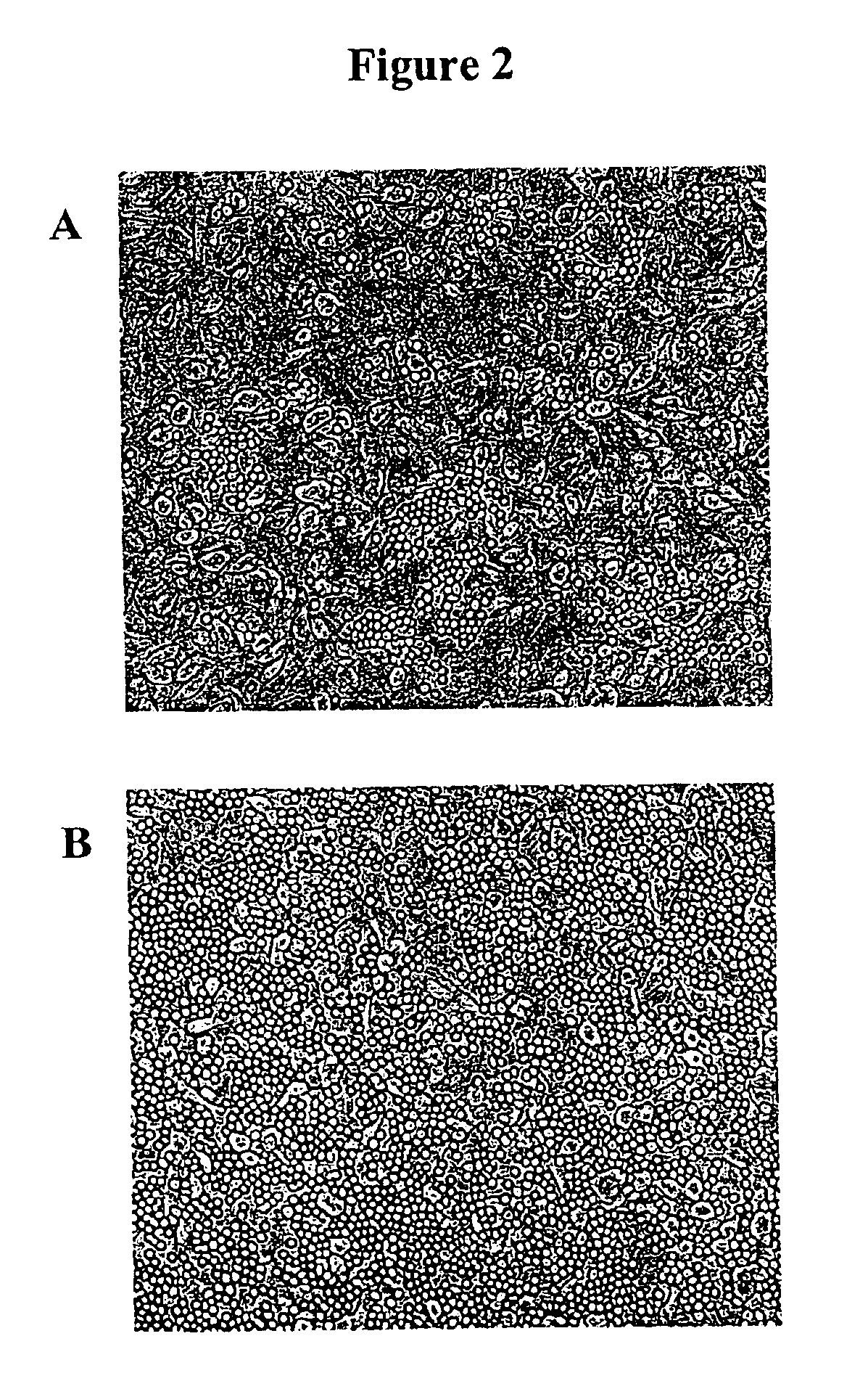
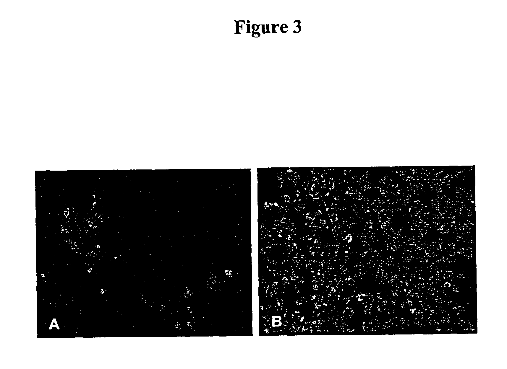
[0088]To compare the sensitivity for the detection of Influenza A by Mv1Lu-hF cells with the parental Mv1Lu cells, the hemadsorption (HAD) and immunofluorescence staining of infected cells was determined as follows. Briefly, each cell type was seeded at a density of 2×10⁵ cells / ml onto a 48-well plate using 0.2 ml per well. After the cells formed a monolayer (usually about 2-3 days), 17 previously tested influenza A virus-positive clinical samples from frozen stocks were inoculated into two wells of each cell with an inoculum of 5 μL / well. After overnight incubation at 36° C. in a 5% CO2 atmosphere, the medium was removed and 0.2 ml of guinea pig red blood cells (RBC) was added. The guinea pig RBCs collected in heparin (Charles River) were washed three times in phosphate buffered saline (PBS) and then resuspended at a concentration of 0.2% of RBC, prior to use. After incubating the plates at 4° C. for 30 min, t...
example 3
Production of Influenza A Virus Using Mv1Lu-hF Cells
[0094]Mink lung epithelial cells are an alternative to Madin-Darby canine kidney (MDCK) epithelial cells for the isolation and cultivation of human influenza viruses (See, Schultz-Cherry et al., J. Clin. Microbiol. 36:3718-3720 [1998]; and Huang and Turchek, J. Clin. Microbiol. 38:422-423 [2000]). The results obtained during the development of the present invention and shown in Example 2 above, indicate that mink lung cells expressing human furin (Mv1Lu-hF) are suitable for the production of high titer influenza A virus stocks for vaccine preparations.
[0095]
TABLE 4Comparison of Influenza A Virus TitersVirusH1N1H3N2CellsMv1LuMv1Lu-hFMv1LuMv1Lu-HFDay 12.5 × 1041.0 × 1055.0 × 1045.0 × 104Day 21.5 × 1051.0 × 1075.0 × 1054.0 × 106Day 31.0 × 1052.0 × 1072.5 × 1045.0 × 107
[0096]The transgenic Mv1Lu-hF and parental Mv1Lu cells were plated in 24 well plates and inoculated at a multiplicity of infection of 0.01 with two subtypes of influenza...
PUM
| Property | Measurement | Unit |
|---|---|---|
| volume | aaaaa | aaaaa |
| time | aaaaa | aaaaa |
| composition | aaaaa | aaaaa |
Abstract
Description
Claims
Application Information
 - R&D
- Intellectual Property
- Life Sciences
- Materials
- Tech Scout
- Unparalleled Data Quality
- Higher Quality Content
- 60% Fewer Hallucinations
Browse by: Latest US Patents, China's latest patents, Technical Efficacy Thesaurus, Application Domain, Technology Topic, Popular Technical Reports.
© 2025 PatSnap. All rights reserved.Legal|Privacy policy|Modern Slavery Act Transparency Statement|Sitemap|About US| Contact US: help@patsnap.com


