Method of detecting biological molecules, and labeling dye and labeling kit used for the same

a biological and labeling technology, applied in the field of biological and labeling labeling methods, can solve the problems of high detection cost and insufficient detection sensitivity

Inactive Publication Date: 2006-03-21
SHINCHIRO ISOBE
View PDF23 Cites 8 Cited by
  • Summary
  • Abstract
  • Description
  • Claims
  • Application Information

AI Technical Summary

Benefits of technology

[0008]An object of the present invention is to solve the above-mentioned problems and to provide a highly sensitive method of detecting a biological molecule at lower cost.
[0017]For example, when used as a kit of DNA microarray, a nucleic acid is used as the biological molecule sample and a probe nucleic acid is fixed on a microarray, while the target nucleic acid sample is labeled by reaction with an organic EL-dye, and the labeled target nucleic acid is spotted on the microarray and hybridization can be conducted under this conditions. Further, applying a binding property between avidin (streptavidin) and biotin, the avidin modified with this dye can be used as a biological assay kit for ELISA (enzyme-linked immunosorbent assay), Western blotting and the like. It can also be used as a kit for protein array. A cell can also be stained, due to efficient labeling of biological molecules such as saccharides and proteins.
[0018]According to the present invention, use of an organic EL-dye as a labeling dye for a biological molecule gives the following effects.
[0019]That is, an organic EL-dye shows high quantum yield in solid state (including solid state and semi-solid state) and manifests high fluorescence intensity. Since an organic EL-dye is cheap as compared with Cy3 and Cy5, a biological molecule can be detected at lower cost. Further, an organic EL-dye reacts with a biological molecule almost quantitatively and shows high incorporation ratio, therefore, high detection sensitivity can be obtained. Furthermore, use of said dye increases the degree of freedom of selectivity of fluorescence wavelength, and multiple fluorescence wavelengths of orange, yellow, green, blue and the like can be used. By this, it becomes possible to use two or more fluorescence dyes having large stokes shift (large difference between excited wavelength and fluorescence wavelength), consequently, plural target nucleic acids contained in one sample can also be simultaneously detected. While Cy3 and Cy5 need to be kept in refrigerated state, an organic EL-dye is chemically stable and can be kept for a long time at ambient temperature, therefore, handling thereof is easy.

Problems solved by technology

However, although Cy3 and Cy5 manifest high fluorescence intensity and have a merit of emission even in solid state, they are very expensive, leading inevitably to a highly expensive detection method.
Also, there is a problem that the ratio of incorporation into a sample RNA or DNA is low and sufficient labeling of a sample RNA or DNA is impossible, resultantly, detection sensitivity is not sufficient.

Method used


Image

  • Method of detecting biological molecules, and labeling dye and labeling kit used for the same
  • Method of detecting biological molecules, and labeling dye and labeling kit used for the same
  • Method of detecting biological molecules, and labeling dye and labeling kit used for the same

Examples


synthesis example 1

[0059]A 1,2,5-oxadiazolo-[3,4-c]pyridine derivative was used as the organic EL-dye.

[0060]The scheme for synthesis of an active ester of a 1,2,5-oxadiazolo-[3,4-c]pyridine (hereinafter, abbreviated as EL-OSu) will be shown below.

(1) Synthesis of Diketone Derivative (2)

[0061]In a 500 mL three-necked flask, 37.5 g (0.25 mol) of 4-methoxyacetophenone (1) and 0.15 g of sodium nitrite were dissolved in 100 mL of acetic acid. On a water bath, a solution prepared by dissolving 100 mL of HNO3 in 100 mL of acetic acid was added dropwise over 2 hours. Then, the mixture was stirred at room temperature for 2 days. The reaction mixture was slowly added into 500 mL of water to cause precipitation. The precipitate was filtrated and dissolved in chloroform. The chloroform phase was washed with saturated sodium bicarbonate water, and washed twice with a 10% NaCl aqueous solution. After dehydration over MgSO4, chloroform was distilled off under reduced pressure to obtain 34.5 g (yield: 78%) of oxadia...

synthesis example 2

[0066]An imidazolopyridine ethyl ester derivative was used as a organic EL-dye. The scheme for synthesis of an active ester of a imidazolopyridine ethyl ester (hereinafter, abbreviated as im-EL-OSu) will be shown below.

(1) Hydrolysis of Imidazolopyridine Ethyl Ester (1)

[0067]In a 500 mL three-necked flask, 0.5 g (1.5 mmol) of an ester 1 was dissolved in 50 mL of ethanol. To this was added 0.12 g (2.1 mol) of KOH. After heating to reflux for 5 hours, the reaction mixture was added to 50 mL of water. Into this aqueous solution, concentrated hydrochloric acid was added dropwise to adjust pH to 1 to obtain a precipitate. The precipitate was filtrated and dissolved in chloroform. The chloroform phase was washed with a 10% NaHCO3 aqueous solution and water. Chloroform was distilled off to obtain a residue. The residue was recrystallized from water to obtain 0.3 g (yield: 63%) of a carboxylic acid 2.

(2) Synthesis of Active Ester (3)

[0068]In a 50 mL three-necked flask, 0.2 g (0.6 mmol) of ...

example 1

1. Labeling of Oligonucleotide with Dye

[0069]Labeling of an oligonucleotide with a dye was conducted according to the following Scheme 4.

(Experimental Procedure)

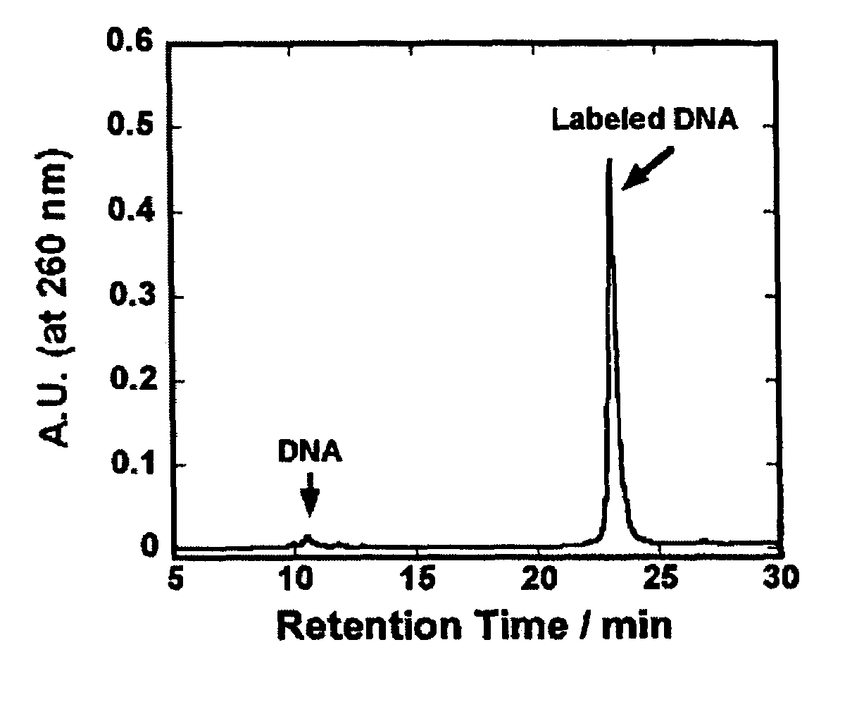
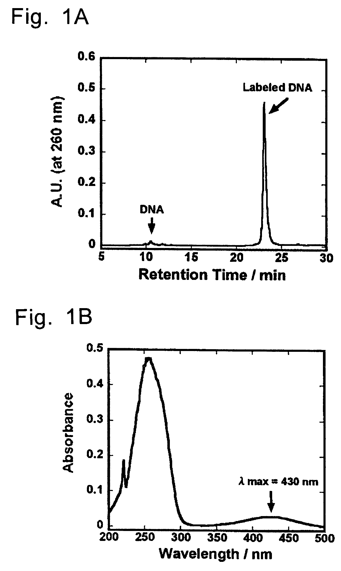
[0070]Into 40 μl of Na2CO3 / NaHCO3 buffer (pH 9.0) containing H2N-dT2O (40 mmol) was added 12 μl of an anhydrous DMSO solution containing 5.0 μmol (2.4 mg) of an active ester of an organic EL-dye and the mixture was shaken at room temperature for 6 hours. After shaking, 0.1 M TEAA (triethylamine acetic acid) buffer (pH 7.0) was added so as to give the total volume of 1 ml, and components derived from the oligonucleotide were separated using NAP-10 column (Pharmacia Sephadex G-25). In this operation, the NAP-10 column had been equilibrated previously with 15 ml of 0.1 M TEAA buffer before use. The sample solution of which total volume had been adjusted to 1 ml was applied into in a column. After elution of 1 ml of the solution, 0.1 M TEAA buffer was charged in a volume of 1.5 ml. Immediately after this, 1.5 ml of the eluted s...


PUM

PropertyMeasurementUnit
temperatureaaaaaaaaaa

Abstract

The present invention provides a method of detecting a biological molecule. The method includes reacting a biological molecule sample with an organic EL-dye and measuring the fluorescence of the biological molecule sample labeled with the organic EL-dye. The method provides a highly sensitive method of detecting a biological molecule at lower cost.

Description

BACKGROUND OF THE INVENTION[0001]1. Field of the Invention[0002]The present invention relates to a method of detecting a biological molecule such as nucleic acids, proteins, peptides, saccharides and the like using a fluorescence dye, and a labeling dye and a labeling kit used for the detection method.[0003]2. Background Art[0004]Recently, post genome researches have been intensively and world widely done aiming for specific gene analysis technologies, gene therapies and tailor made medical treatments. As for the gene analysis technology, a method of detecting DNA using a DNA microarray, for example, is used. According to this detection method, simultaneous analysis of expression, functionality, mutation and the like of a plurality of genes can be conducted simply and quickly.[0005]In the detection method using a DNA microarray, DNA chips obtained by spot-fixing many sequences (probe nucleic acids) of DNA or oligonucleotide on a substrate made of glass, silicon or the like are used....

Claims


Application Information

Patent Timeline
Patent Type & Authority Patents(United States)
IPC IPC(8): C12Q1/68C07H21/00C07H21/02C07H21/04G01N33/58C07F5/02C09B57/00C09K11/06
CPCC07D471/04C07D498/04C07H21/02G01N33/582C07H21/04C12Q1/6816C09B57/00C09K11/06C09K2211/1044C09K2211/1048C12Q2563/107C12Q2563/113
Inventor ISOBE, SHINICHIRO
Owner SHINCHIRO ISOBE
Why Eureka
Try Eureka