Transgenic luciferase mouse

a luciferase mouse and mouse technology, applied in the field of transgenic animals, can solve the problems of pet being regarded as an expensive test, small tumors or tumors in difficult to reach areas can go undiagnosed by palpation, and cumbersome animal models

Inactive Publication Date: 2006-05-09
SLOAN KETTERING INST FOR CANCER RES
View PDF0 Cites 2 Cited by
  • Summary
  • Abstract
  • Description
  • Claims
  • Application Information

AI Technical Summary

Benefits of technology

[0019]FIG. 7 shows that a readout of therapeutic effect is not dependent on the size of the tumor being treated. Several mice with PDGF-induced gliomas having various light outputs were treated with PTK787. Bioluminescence output was quantified daily during treatment. The log and the percent graph show essentially parallel responses of decreases in light emission over time with treatment.

Problems solved by technology

However, animal models traditionally have been cumbersome because of the difficulty in quantitating tumor burden and the requirement for either bulk tumor growth or animal survival as end points to evaluate the effect of a potential therapy.
Small tumors or tumors in difficult to reach areas can go undiagnosed by palpation and can be refractory to caliper measurements.
However PET is regarded as an expensive test, demanding technique that is time consuming and substantial expertise and training with a considerable infrastructure (cyclotron, supporting radiochemistry laboratory space, usually two PET scanners, and a significant number of support staff).
These reasons make this method too cumbersome and expensive for analysis of large numbers of animals in an experimental therapy evaluation.
However, even though MRI does not need radioactive isotopes, it does require expensive equipment and time consuming analysis of the data, making it less than adequate for high throughput analysis of animal models.
Transplanted tumors are, however, only a partial model of human tumorigenesis because the histology of these tumors does not resemble that of the human disease.
Moreover, because these tumor models have not been predictive in preclinical trials, the biology of these tumors may not recapitulate the human disease.
Specific bioluminescence tumor models are by its own nature of limited use as a tool for the general study of many different types of cancer in animal models.

Method used


Image

  • Transgenic luciferase mouse
  • Transgenic luciferase mouse
  • Transgenic luciferase mouse

Examples


example 1

Generation of Elux Transgenic Mice

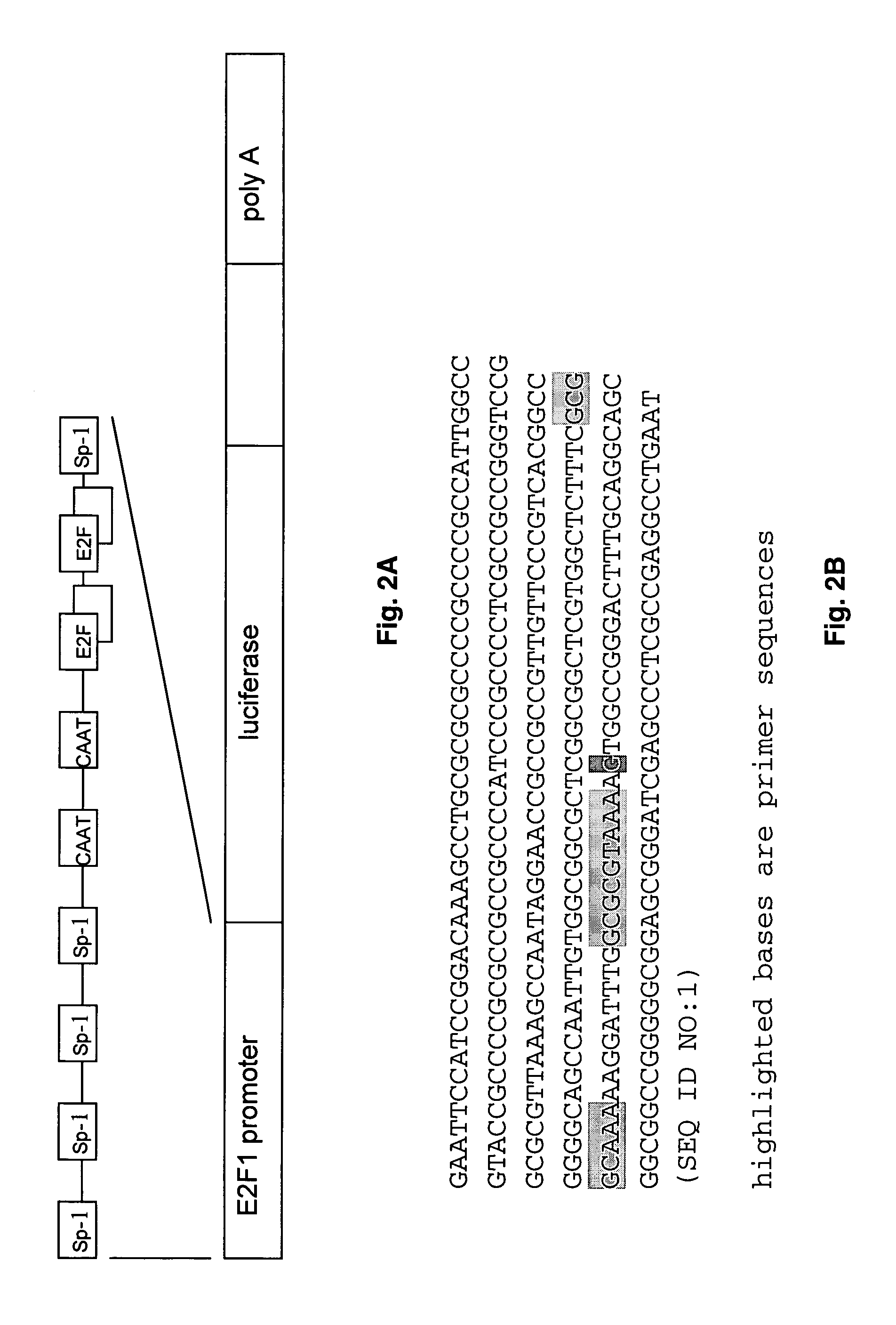
[0037]The Elux reporter transgene was constructed by ligation of the E2F1 promoter obtained from David Johnson (MD Anderson Cancer Center) with the firefly luciferase as illustrated in FIG. 2. The E2F1 promoter used includes 208 nucleotides upstream, and 66 bases downstream of the transcription site. The sequence contains several binding sites for E2F1 as well as binding sites for the transcription factor SP1. This was digested with EcoR1 and Stu1 and ligated to the gene encoding firefly luciferase digested with compatible restriction enzymes. These fragments were incubated with DNA ligase and used to transform E. coli. The plasmid DNA was then purified and characterized by sequence analysis. This analysis indicated a point mutation in the E2F1 promoter that resulted in a change in the sequence relative to the published sequence as indicated in FIG. 2. The mutation indicated did not affect the binding sites for any known transcription factors, and t...

example 2

The Use of Elux Transgenic Mouse as a Glioma Animal Model

[0038]The following examples demonstrate Elux transgenic mouse can be used to monitor the formation of tumors in genetically defined tumor models such as germline modification or somatic cell gene transfer technologies.

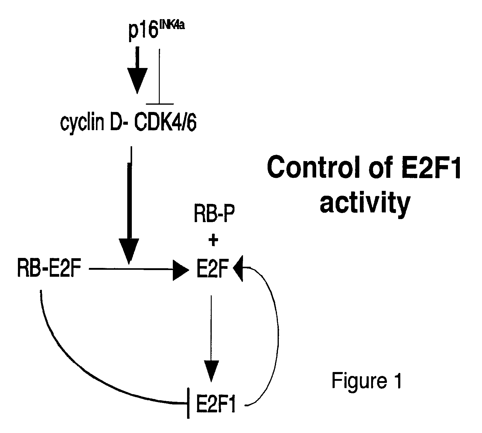
[0039]Germline modification models of tumor formation such as transgenic mice or mice with targeted deletions, conditional knockouts, and inducible systems can be crossed into the Elux transgenic background. When these models form tumors, the deregulation of the Rb pathway is identified as the expression of the Elux transgene and detectable by bioluminescence. This allows non-invasive monitoring of tumor presence and activity in response to therapeutic and genetic intervention.

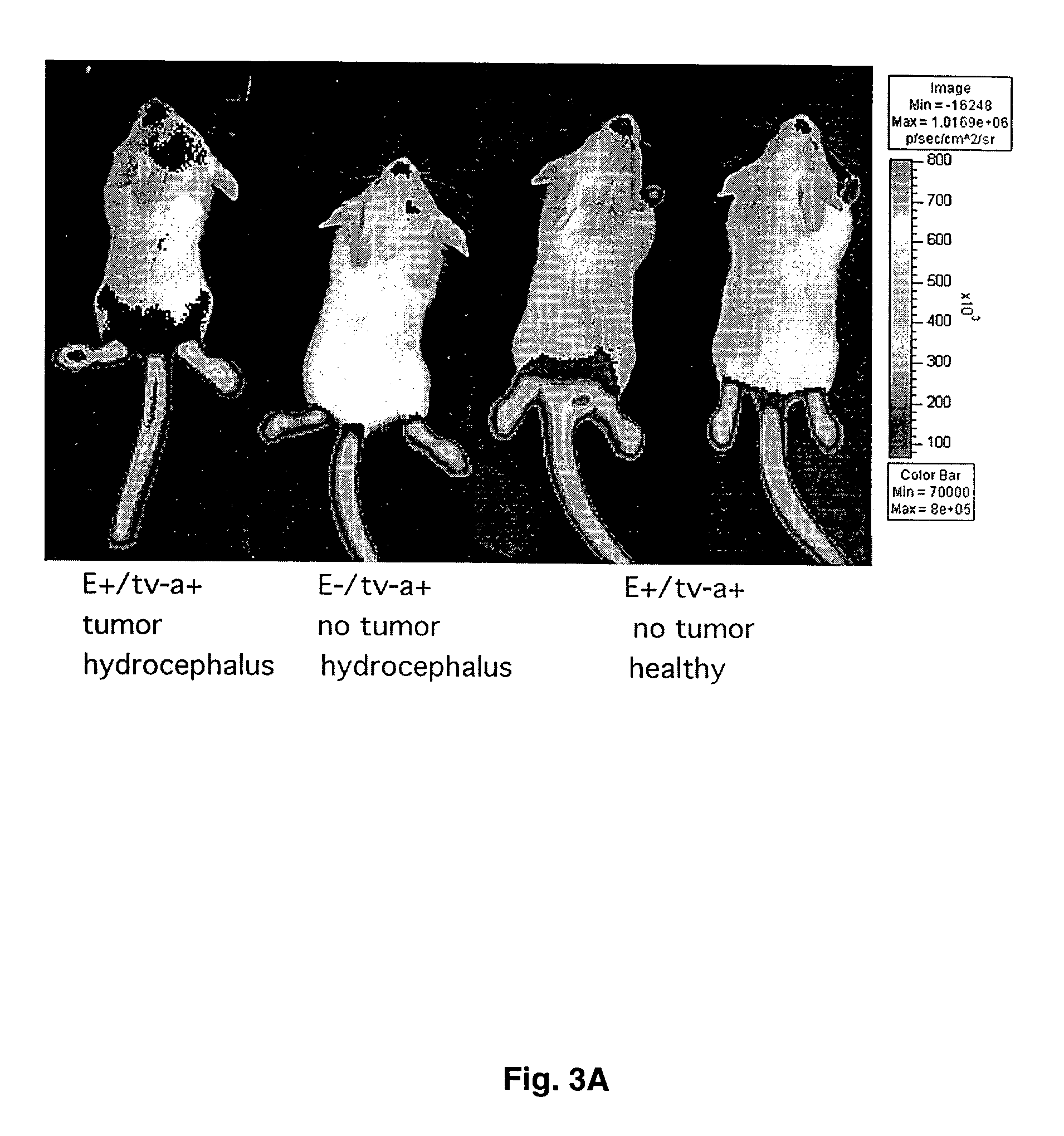
[0040]As an illustration of the use of Elux in somatic cell gene transfer models, the inventors have generated gliomas in mice harboring the Elux transgene. The Elux transgenic line was crossed with the Ntv-a transgenic mouse line. These d...

example 3

In Vivo Imaging of Tumors

[0041]Animals were weighed prior to the acquisition session for proper dosage calculations. A fresh sterile solution of D-Luciferin (Xenogen, XR-1001) was prepared in the following dosage: 150 mg / kg D-Luciferin in 3 ml / kg Normal Saline. The solution was sterilized by filtration through 0.22 μm syringe filter. Individual dosages were drawn into sterile insulin syringes for each animal based on the body weight. Prior to the injection, individual syringes were kept in dark. During all the procedures D-Luciferin was protected from light.

[0042]Inhalation anesthesia machine was connected to the gas inlet hose of the IVIS. Gas inlet selector was turned into the “gas on” position. The outlet hose has to be connected to the central vacuum system and the valve open for a low flow active aspiration of the chamber air to eliminate exposure of the operator to the inhalation gas mixture and to avoid the animals' overexposure to the anesthetic. The inhalation anesthesia ma...


PUM

PropertyMeasurementUnit
red wavelengthsaaaaaaaaaa
temperatureaaaaaaaaaa
weightaaaaaaaaaa

Abstract

The present invention provides a transgenic animal expressing the reporter gene, luciferase, driven by a promoter (e.g. the E2F1 promoter) that acts as a sensor of cell cycle. The luciferase substrate, luciferin, emits light when metabolized, and the light is transmitted through mammalian tissues. Therefore, the transgenic animal model of the present invention allows for monitoring of areas of major cell cycle activity, a characteristic of cancer cells, under adequate visualization conditions. These transgenic animals are useful as in vivo models for testing preventative measures for cancer as well as for testing novel therapeutic modalities.

Description

CROSS-REFERENCE TO RELATED APPLICATION[0001]This non-provisional patent application claims benefit of priority of provisional patent application No. 60 / 416,001 filed Oct. 4, 2002, now abandoned.BACKGROUND OF THE INVENTION[0002]1. Field of the Invention[0003]The present invention relates to the use of a transgenic animal for in vivo monitoring of animal models of human cancers. The transgenic animal is useful for testing potential preventative measures and therapeutic modalities as well as determining causative agents of cancer.[0004]2. Description of the Related Art[0005]The study of the molecular mechanisms of tumorigenesis has been greatly facilitated in recent years by the use of animals able to overexpress or not, at the tissue specific or general level, a variety of genes (germ line modification). However, animal models traditionally have been cumbersome because of the difficulty in quantitating tumor burden and the requirement for either bulk tumor growth or animal survival as...

Claims


Application Information

Patent Timeline
Patent Type & Authority Patents(United States)
IPC IPC(8): A01K67/00A01K67/027A01K67/033C07H21/04C12NC12N15/00C12N15/85G01N33/00
CPCA01K67/0275C12N15/8509A01K2217/05A01K2227/105C12N2830/007A01K2267/0393A01K2267/0331
Inventor HOLLAND, ERIC CHARLES
Owner SLOAN KETTERING INST FOR CANCER RES
Why Eureka
Try Eureka