Method and device for trichomonas detection

a technology of trichomonas and kit, which is applied in the field of immunoassay method and kit for detecting trichomonas infection, can solve the problems of adversely affecting both fertility and pregnancy, increasing the risk of acquiring cervical cancer, and severe health effects of infection with trichomonas

Inactive Publication Date: 2007-11-06
XENOTOPE DIAGNOSTICS INC +1
View PDF27 Cites 7 Cited by
  • Summary
  • Abstract
  • Description
  • Claims
  • Application Information

AI Technical Summary

Benefits of technology

[0028]In one aspect, the invention includes a method for detecting Trichomonas vaginalis infection in a human subject. In practicing the method, a body-fluid sample, such as a urine or vaginal-swab sample, is obtained from the subject and contacted with an antibody specific against a Trichomonas adhesin peptide, to form an antibody-adhesin peptide complex if the subject is infected with Trichomonas. The presence or absence of the complex is diagnostic with a reliability of at least about 80%, and typically beterr than 90%, where the clinical specimen is a vaginal swab, and with a reliability of higher than 40% where the clinical specimen is urine.

Problems solved by technology

In humans, infection with Trichomonas has severe health consequences.
Moreover, infection by Trichomonas increases the risk of acquiring cervical cancer, and can adversely affect both fertility and pregnancy (Yap et al., 1995; Zhang et al., 1995).
Wet mount microscopy is highly inefficient when performed on urine samples, with sensitivity as low as 7% (compared to culture at 40%) (van Der Schee C., et al. J. Clin Microbiol.
In addition, the wet mount microscopy method of detection, utilizing any of the sample types described, is subjective, tedious and time consuming, and requires a trained person.
Unfortunately, the procedure requires several days to complete (typically 2-5 days) and must be conducted in special laboratories with trained personnel.
Further, both of these methods require the parasites in the samples to be viable before they can be detected, constraining the suitability of certain sample types.
For example, it has been determined that urine is not a suitable sample medium for culture-based diagnosis of Trichomonas, despite being more sensitive than wet mount (Mohamed et al.
Although PCR is potentially more sensitive than wet mount or culture (87% according to the above reference), there is a lack of scientific consensus on what primer sequences are Trichomonas specific (PCR is not an FDA approved diagnostic method).
Further, the problem of false positives in both discrepant samples and contaminated clinical sampleshinders the application of this technique.
In addition, PCR is a highly technical procedure with many manipulations and procedural controls, requires expensive equipment, and does not allow for point of care diagnosis; all factors mitigating against its suitability as a practical diagnostic method for Trichomonas.
The method involves a large number of manipulations and the use of expensive detection equipment, and is less sensitive that wet mount microscopy.

Method used


Image

  • Method and device for trichomonas detection
  • Method and device for trichomonas detection
  • Method and device for trichomonas detection

Examples


example 1

Purification of Monoclonal Antibody

[0115]Monoclonal anti-Trichomonas antibodies (Mabs) were prepared as described in U.S. Pat. No. 4,707,442 to Alderete. The Mabs were purified either from culture supernatant or ascites by protein A-Sepharose chromatography (Goding, J Immunol Meth (1976) 42:17) (Pharmacia LKB). About 2 to 6 mL of ascites fluid produced from the appropriate hybridoma was diluted 1:1 with Binding Buffer containing 1.5M glycine, 3 M NaCl adjusted to pH of about 8.9. The DM116 Mab ascities were subjected to dialysis with binding overnight at room temperature. The dialysed Mab ascites were eluted from the protein A-Sepharose CL-4B affinity gel chromatography column with the Binding Buffer at a rate of 0.5 mL / minute. Elution was monitored by determining the absorbance of each sample at 280 nm, and the column was washed until the absorbance was below 0.001 O.D. Then, IgG was eluted from the column using 0.1 M citric acid buffer adjusted to an approximate pH 4.0. Elution of...

example 2

Preparation of Monoclonal Antibody Gold Conjugate

[0116]A flask containing 100 mL of ultra-pure water was heated to about 80-85° C. Then, with gentle stirring, 1 mL of 1% AuCl (2×0.5 mL) and 1.75 mL of 1% sodium citrate solution (2×0.5, and then 0.75 mL aliquots) were consecutively added to the hot water. The solution was further heated at 80-85° C. until the color changes from slightly yellow to purplish black to cherry wine.

[0117]In a glass tube, to 10 mL of the 1:1.75 gold solution prepared as above was added 200 μL of freshly prepared 100 mM of carbonate (100 mM). The solution was vortexed for 3 seconds. Then 300 μL of DM116 IgG (1 m / mL) was added, and the solution was vortexed for 3 seconds again. The tube was incubated for 30 min. at room temperature, 100 μL of the conjugate blocking solution was added with vortexing for 3 seconds, followed by centrifugation at 8000 rpm for 30 min. The centrifuge was allowed to come to a complete stop without the use of the breaking system. The...

example 3

Preparation of Assay Dry Strip

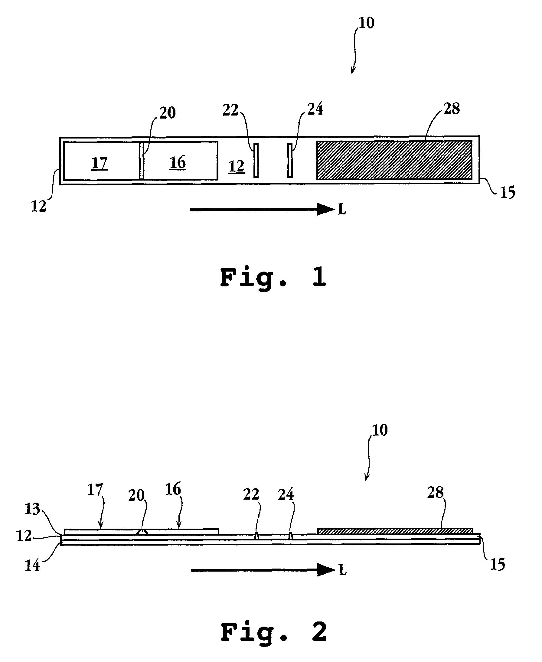
A. Addition of Immobilized Antibodies to Dry Strip

[0118]A preparation of 1 mg / mL of the rabbit anti-T. vaginalis IgGs was dialyzed overnight at room temperature against the coating buffer consisting of 50 mM carbonate-bicarbonate buffer at pH 9.6. The IgGs were then loaded onto the X-Y-Z Dispensing Platform (BIO.DOT INC.), and the machine was programmed to deliver 1 μL / cm of the liquid onto a nitrocellulose membrane. The membrane was then dried at room temperature to 37° C. for about 3 h. The blocking buffer (0.5% BSA; 4% sucrose in PBS) was then sprayed over the membrane, and the membrane was dried overnight at room temperature. The membrane was stored at about 2-8° C. in the absence of moisture.

B. Application of gold Conjugate DM116 Antibody at the Reaction Zone

[0119]A glass fiber filter paper was submersed into the gold pad pre-treatment buffer (0.5%(w / v) poly alcohol; 0.71% (w / v) di-sodium phosphate; 0.1% (v / v); Triton X-100; 0.5% BSA (w / v); DDW; pH...


PUM

PropertyMeasurementUnit
volumeaaaaaaaaaa
temperatureaaaaaaaaaa
widthaaaaaaaaaa

Abstract

A method and kit for detecting Trichomonas vaginalis infection in a human subject are disclosed. In the method, a body-fluid sample such as a vaginal swab sample or urine is obtained from the subject and contacted with an antibody specific against a Trichomonas adhesin peptide, forming an antibody-adhesin peptide complex if the subject is infected with Trichomonas. The presence or absence of the complex establishes, with a reliability of at least 80%, in the case of a vaginal swab sample, and with a reliability of at least 40% in the case of a urine sample, the presence or absence, respectively, of Trichomonas infection in the subject. A preferred test kit employs a dry-strip, sandwich assay, format.

Description

[0001]The subject application claims priority to Provisional Application, Ser. No. 60 / 302,733, filed on Jul. 3, 2001, entitled Diagnostic Tests and Kits for Trichomonas.FIELD OF THE INVENTION[0002]The present invention relates to an immunoassay method and kit for detecting Trichomonas infection.REFERENCES[0003]Alderete et al., Infect. Immun., 40:284-291, 1983[0004]Alderete et al., Infect. Immun., 49:463-468, 1985.[0005]Alderete et al., Infect. Immun., 52:70-75, 1986.[0006]Alderete et al., Infect. Immun., 53:285-293, 1986.[0007]Alderete et al., Infect. Immun., 53:697-699, 1986.[0008]Alderete et al., Infect. Immun., 55:1037-1041, 1987.[0009]Alderete and Garza, Infection and Immunity, 56(1):28-33, 1988.[0010]Alderete and Garza, Infection and Immunity, 56(10):2256-2562, 1988.[0011]Alonzo and Pepe, Statistics in Medicine, 18:2987-3000, 1999.[0012]Arroyo et al., Molecular Microbiology, 6(7):853-862, 1992.[0013]Arroyo et al., Arch Med Res 26(4):36-369, 1995.[0014]Arroyo et al., Molecular M...

Claims


Application Information

Patent Timeline
Patent Type & Authority Patents(United States)
IPC IPC(8): G01N33/554G01N33/543G01N33/558G01N33/571G01N33/569G01N33/58
CPCG01N33/558G01N33/56905G01N2333/44G01N33/543G01N33/53
Inventor ALDERETE, JOHN P.CASTELLA, PAUL C.
Owner XENOTOPE DIAGNOSTICS INC
Why Eureka
Try Eureka