Screening method for developing drugs against pathogenic microbes having two-component system

a technology of pathogenic microorganisms and screening methods, applied in the field of screening methods, can solve the problems of no new intervention reported, no functional dissection of the mycobacterial regulatory network cascade involving a two-component system in general, and the devr-devs system in particular

Inactive Publication Date: 2009-07-28
COUNCIL OF SCI & IND RES
View PDF3 Cites 3 Cited by
  • Summary
  • Abstract
  • Description
  • Claims
  • Application Information

AI Technical Summary

Problems solved by technology

However, functional dissection of a mycobacterial regulatory network cascade involving a two-component system in general, and DevR-DevS system in particular, has not been accomplished yet.
thers. Despite such a huge number of genes targeted for anti-tubercular therapy, no new interventions have been reported t
But, incremental changes in the structure of poorly efficacious antibacterials, however, are likely to afford analogues with a limited life span due to established resistance to the parent drug (Macielag and Goldschmidt, 2000).
However, to date such a strategy has not been attempted for developing antimycobacterial agents.
However, the precise mode of action of such inhibitors has generally remained obscure due to lack of published enzyme kinetics data, making it difficult to develop a useful pharmacophore model.
Drug resistant forms of tuberculosis pose a serious threat to the successful outcome of a tuberculosis control programme in a community.
Although many highly effective drugs such as isoniazid, rifampicin, ethambutol and pyrazinamide are available, poor compliance due in part to long treatment schedules (6 months is the standard duration) leads to high rates of treatment failure.
Though conventional drug regimens comprising administration of rifampicin-isoniazid-pyrazinamide is very effective, the minimum inhibitory concentration (MIC) for all the three drugs is very close to the maximum serum concentration, which is limited by toxicity, resulting in poor therapeutic index for each.
This ineffectiveness of current therapies is therefore directly responsible for both the very long duration of therapy and the emergence of resistance to drugs.
Certain fourth generation quinolones including gatifloxacin and levamofloxacin etc. have shown certain degree of efficacy against many—a clinical isolates of mycobacteria including M. tuberculosis under in vitro conditions, but these drugs are also marred by the same intrinsic drawbacks like their limited efficacy under in vivo conditions, severe side-effects, high cost and rapid emergence of resistant isolates.
Although many lead compounds targeted against the conventional drug targets are being tested worldwide, no new drugs for tuberculosis have been introduced in the market over the last thirty years.
In addition, there is no drug available in the market today which can be used to tackle the ever expanding problem of latent tuberculosis.
All the drugs which are used for combating tuberculosis, are effective only in the active disease conditions and are not able to eradicate the latent disease.
But, all the identified inhibitors and compounds for bacterial two-component systems suffer from the drawback of extreme hydrophobicity.
Such high hydrophobicity of these molecules makes formulation and delivery of the compounds extremely difficult.
Furthermore, the compounds showed minimal bioavailability and excessive plasma protein binding, Thus, the compounds have been ineffective in standard in vivo infection models.
Though these compounds demonstrated good antibacterial activity, they also suffered from excessive protein binding, which hampered in vivo efficacy.
But, the mechanism of cell death was shown to be membrane-disruption leading to complete shutdown of macromolecular synthesis.
The compounds are also ineffective in vivo due to excessive plasma protein binding.
Thus far, all identified inhibitors of autophosphorylation of histidine sensor kinases have turned out to be hydrophobic compounds that were often difficult to formulate adequately for in vivo protection studies.
Whenever tested, these compounds failed to protect in mouse models of infection probably due to excessive serum protein binding.
However, isothiozolone derivatives had already shown their inefficacies in in vivo studies as mentioned above.
However, as mentioned above the imidazolium derivatives and analogs clearly have a general effect on histidine protein kinase autophosphorylation as well and are quite ineffective in in vivo protection assays.
In general, most of these agents appear to suffer from poor selectivity, excessive protein binding or limited bioavailability.

Method used


Image

  • Screening method for developing drugs against pathogenic microbes having two-component system
  • Screening method for developing drugs against pathogenic microbes having two-component system
  • Screening method for developing drugs against pathogenic microbes having two-component system

Examples


example 1

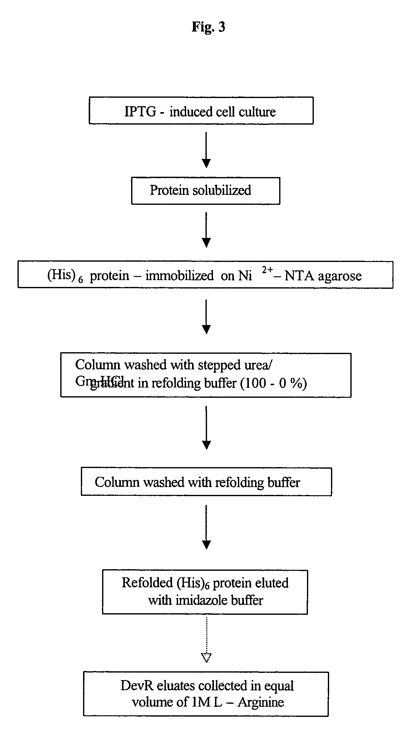
Cloning, Overexpression and Purification and Refolding of DevR, DevS and Rv2027c and Their Single Domain Derivative / s and Mutant Variant Proteins

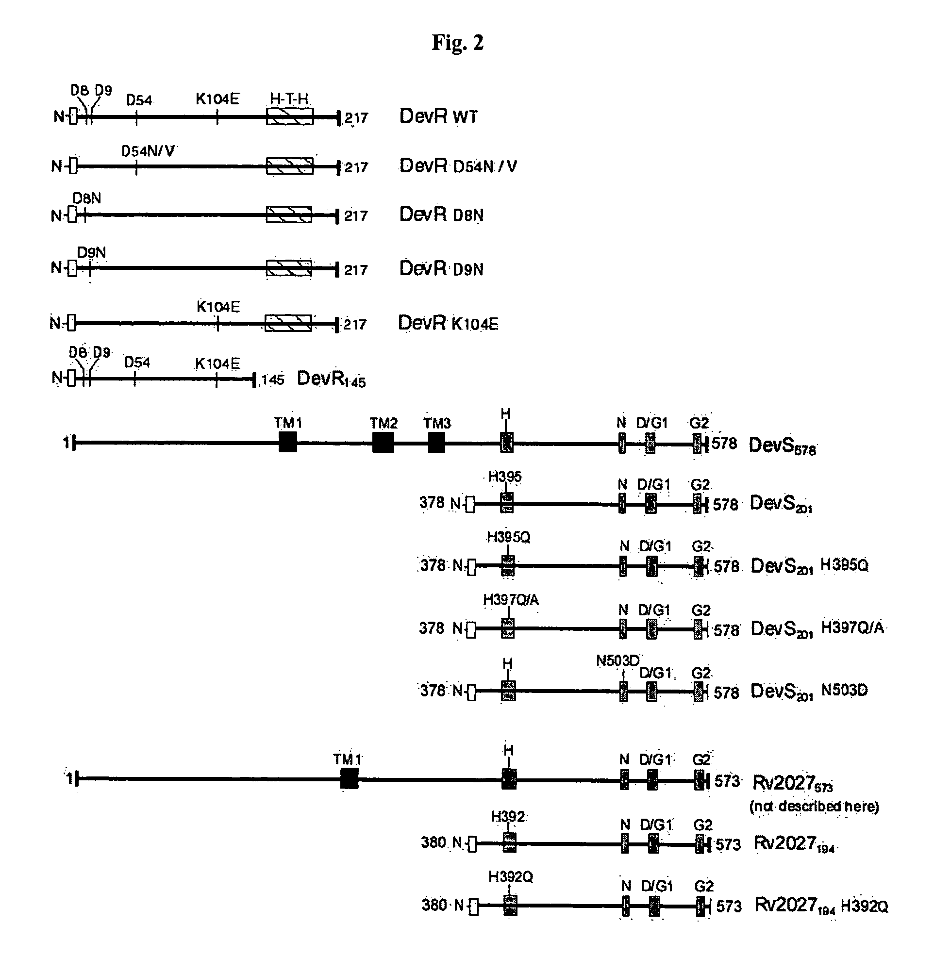
A. Cloning of DevR, DevRN145, DevS201, DevS578 and Rv2027194 and Generation of Their Single Domain and Mutagenic Derivatives

[0134]All routine recombinant DNA work was performed as described (Sambrook and Russell, 2001). M. tuberculosis H37Rv DNA was prepared by boiling cells in the presence of 0.1% Triton X-100 for 25 minutes at 95° C. and the supernatant recovered after centrifugation was used as a source of template DNA in PCR. Full length devR, N-terminal truncated devS201, full length devS578 and N-terminal truncated Rv2027194 genes were amplified from M. tuberculosis DNA by PCR using Pfu DNA polymerase and primers that contain restriction enzyme sites engineered in them (FIG. 2, Table 1).

[0135]For the expression of N-terminally His6-tagged DevR protein, the devR PCR product was digested with NdeI and SalI and after blunting with T4 DNA...

example 2

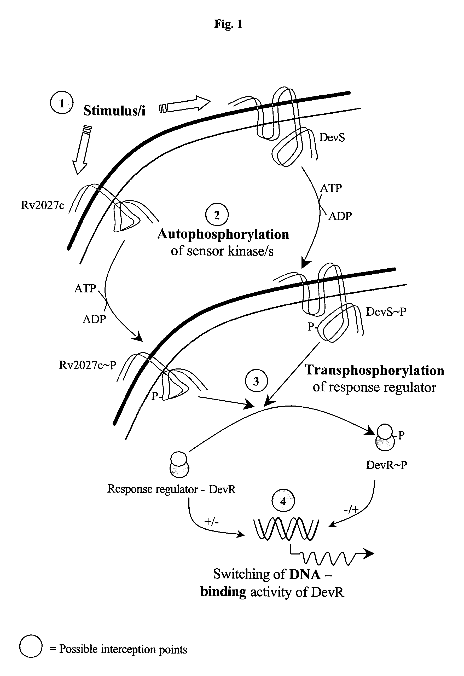
Studies of Autophosphorylation Activity of DevS and Rv2027c Proteins

[0163]DevS201, DevS578 and Rv2027194 proteins and their mutant derivatives DevS201-H395Q, DevS201-H397Q, DevS201-H397A, DevS201-N503D and Rv2027194-H392Q were subjected to autophosphorylation with [α32P] ATP as phosphodonor molecule.

[0164]Purified and refolded protein / s (15 μM of DevS201 or Rv2027194 or their mutagenic variants thereof and 2.5 μM of DevS578) were incubated with 5 μCi of [α32P]ATP (5000 Ci / mmol, BRIT, CCMB campus, Hyderabad, India) in 10 μl reaction buffer (50 mM Tris.HCl, pH 8.0, 50 mM KCl, 10 mM MgCl2, 50 μM ATP) at 25° C. for the designated periods of time. After which the reactions were terminated by addition of 5 μl 3× sample buffer containing 250 mM Tris.HCl, pH 6.8, 10% glycerol, 1% SDS, 280 mM β-mercaptoethanol, 0.01% bromophenol blue.

[0165]The samples were immediately frozen on chilled ethanol bath and stored at −70° C. until just prior to analysis by SDS-PAGE.

[0166]Prior to SDS-PAGE analysi...

example 3

Phosphorylation Activity of DevR

[0174]To study the phosphotransfer reaction, DevS201 / Rv2027194 (15 μM) or DevS578 (5 μM) was phosphorylated for 30 min as described above. Subsequently, DevR / DevRN145 protein (20 μM) was added to 32P phosphorylated sensor kinases. At indicated time points, aliquots were removed and stored at −70° C. until analysis. Subsequently, the samples were analysed on 15% SDS-PAGE and subjected to autoradiography as described for autophosphorylation reactions.

[0175]The phosphorylation assays with the DevRN145 and DevR mutant proteins were performed under identical conditions as described for the wild type DevR protein. The findings are provided below:[0176]1. Phosphorylation of DevR / DevRN145 by phosphotransfer from DevS578, DevS201 and Rv2027194 occurred very rapidly. Within 2-5 minutes of the initiation of reaction, half of the radioactivity incorporated in the sensor kinases was transferred to DevR (FIGS. 9A, B, C, D, E). These rapid kinetics would be useful f...


PUM

PropertyMeasurementUnit
volumeaaaaaaaaaa
pHaaaaaaaaaa
pHaaaaaaaaaa

Abstract

The present invention relates to a package of screening methods for developing drugs against pathogenic microbes having two-component system of DevR-DevS and / or DevR-Rv2027c and its homologues, said method comprising steps of over-expressing DevR, DevS, and Rv2027c and their single domain derivatives including mutant variant proteins, autophosphorylating DevS, and Rv2027c proteins and thereafter, phosphotransfering to DevR and its derivatives in SDS-PAGE or High-throughput format in the presence of a test compound, and determining the drug-potential of the test compound, wherein the potential of the drug is inversely proportional to (i) the degree of autophosphorylation of DevS and Rv2027c, (ii). the degree of phosphotransfer-based dephosphorylation of DevR and / its single domain derivative, and (iii). the degree of dephosphorylation of phosphorylated species of DevS and Rv2027c and / their single domain derivatives, and a method of treatment, and a composition thereof.

Description

SPECIFICATION[0001]The present application claims priority to U.S. Provisional Application Ser. No. 60 / 418,837, filed Oct. 16, 2002, which is incorporated herein by reference in its entirety.FIELD OF THE PRESENT INVENTION[0002]The present invention relates to screening methods designed for identifying drugs for use against pathogenic microbes. The screening method of the invention is a two-component system of DevR-DevS and / or DevR-Rv2027c and its homologues, wherein said method comprises the steps of over-expressing DevR, DevS, and Rv2027c and their single domain derivatives including mutant variant proteins, autophosphorylating DevS, and Rv2027c proteins and thereafter, phosphotransfering to DevR and its derivatives in SDS-PAGE or High-throughput format in the presence of a test compound, and determining the drug-potential of the test compound, wherein the potential of the drug is inversely proportional to (i) the degree of autophosphorylation of DevS and Rv2027c, (ii) the degree o...

Claims


Application Information

Patent Timeline
Patent Type & Authority Patents(United States)
IPC IPC(8): C12Q1/48A61K45/06A61P31/04C12Q1/18G01N33/68
CPCA61K45/06C12Q1/18G01N33/6893C12Q1/485G01N2500/00A61P31/04
Inventor TYAGI, JAYA SIVASWAMISAINI, DEEPAK KUMAR
Owner COUNCIL OF SCI & IND RES
Why Eureka
Try Eureka