Oral-intestinal vaccines against diseases caused by enteropathic organisms using antigens encapsulated within biodegradable-biocompatible microspheres

a biocompatible microsphere and enteropathogenic technology, applied in the field of oral-intestinal vaccines against enteropathogenic organisms using biodegradable biocompatible microspheres, can solve the problems of time-consuming and costly experiments to identify these epitopes, and the effective method of parenteral immunization to induce mucosal immunity is not effective, so as to prevent intestinal bleeding, enhance the immunogenicity of antigens, and promote intestinal bleeding. mild but significant

Inactive Publication Date: 2009-10-20
UNITED STATES OF AMERICA THE AS REPRESENTED BY THE SEC OF THE ARMY
View PDF50 Cites 0 Cited by
  • Summary
  • Abstract
  • Description
  • Claims
  • Application Information

AI Technical Summary

Benefits of technology

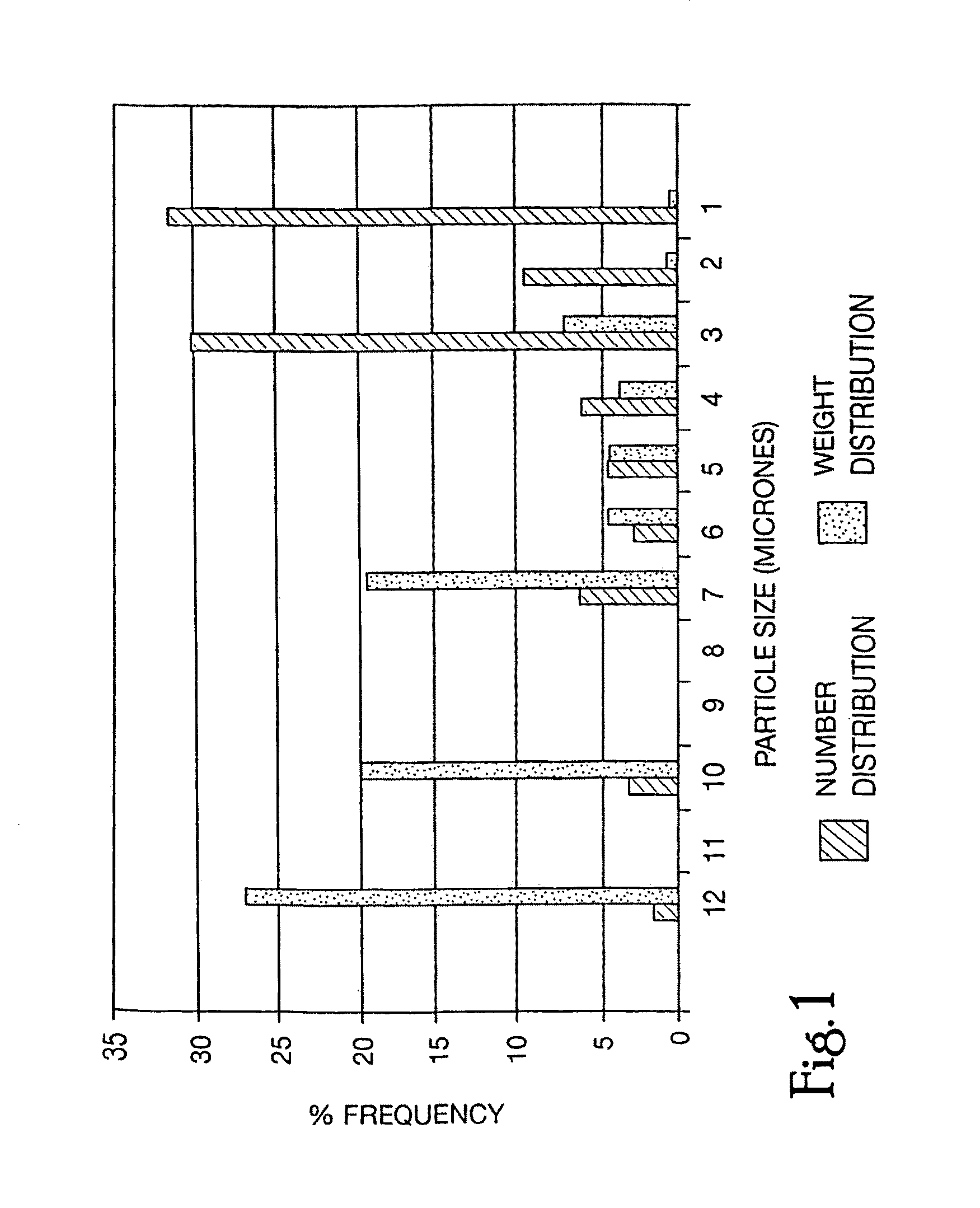
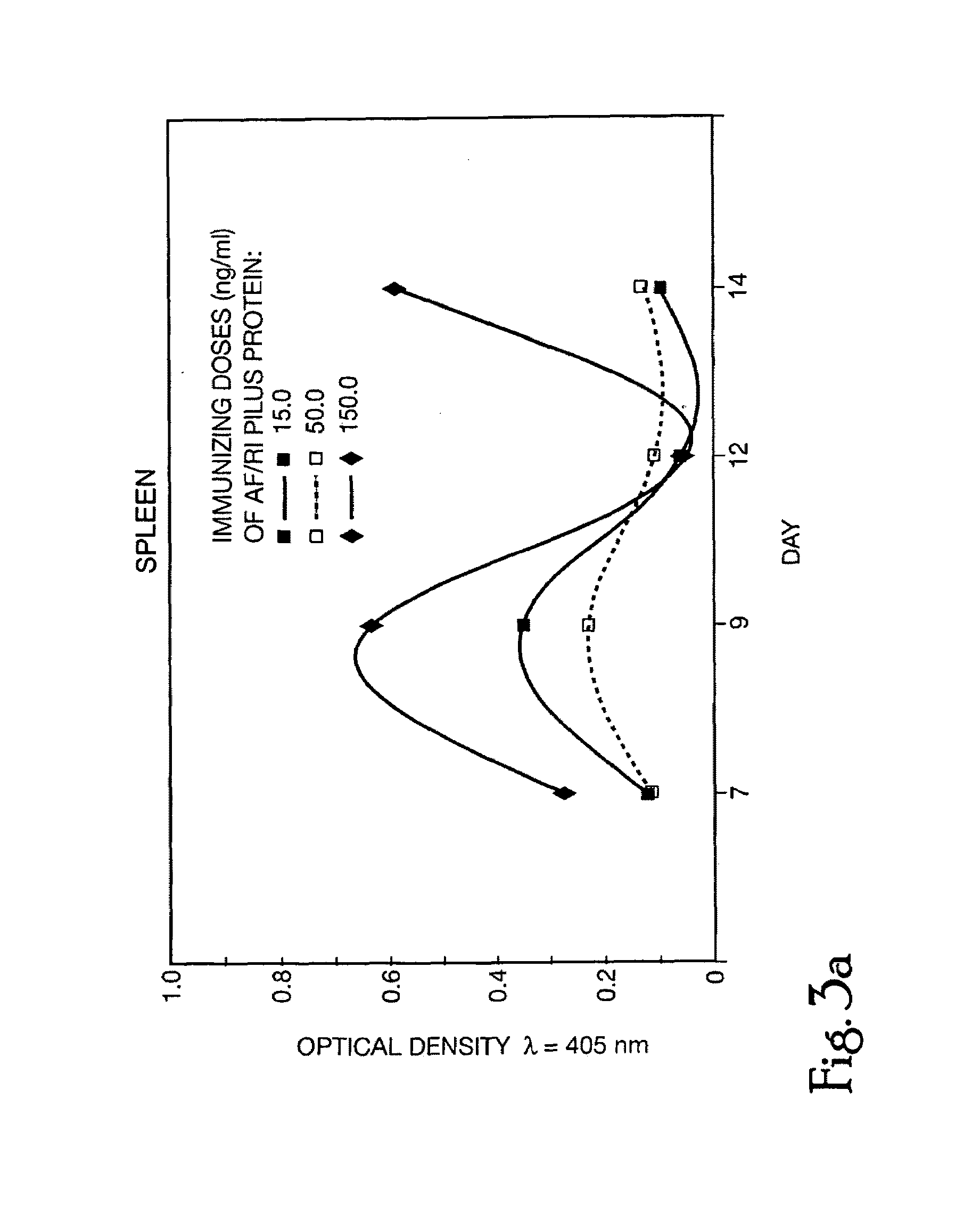
[0010]This invention relates to a novel pharmaceutical composition, a microcapsule / sphere formulation, which comprises an antigen encapsulated within a biodegradable polymeric matrix such as poly (DL-lactide-co-glycolide) (DL-PLG), wherein the relative ratio between the lactide and glycolide component of the DL-PLG is within the range of 40:60 to 0:100, and its use, as a vaccine, in the effective pretreatment of animals (including humans) to prevent intestinal infections caused by a virus or bacteria. In the practice of this invention, applicants found that the AF / R1 adherence factor is a plasmid encoded pilus composed of repeating pilin protein subunits that allows E. coli RDEC-1 to attach to rabbit intestinal brush borders. To identify an approach that enhances the immunogenicity of antigens that contact the intestinal mucosa, applicants investigated the effect of homogeneously dispersing AF / R1 pili within biodegradable microspheres that included a size range selected for Peyer's Patch localization. New Zealand White rabbits were primed twice with 50 micrograms of either microencapsulated or nonencapsulated AF / R1 by endoscopic intraduodenal inoculation. Lymphoid tissues were removed and cellular proliferative responses to AF / R1 and synthetic AF / R1 peptides were measured in vitro. The synthetic peptides represented possible T and / or B cell epitopes which were selected from the AF / R1 subunit sequence using theoretical criteria. In rabbits which had received nonencapsulated AF / R1, Peyer's Patch cells demonstrated slight but significant proliferation in vitro in response to AF / R1 pili but not the AF / R1 synthetic peptides. In rabbits which had received microencapsulated AF / R1, Peyer's Patch cells demonstrated a markedly enhanced response to AF / R1 and the synthetic peptides. Cells from the spleen and mesenteric lymph nodes responded similarly to AF / R1 pili in both groups of animals, while there was a greater response to the synthetic peptide AF / R1 40-55 in rabbits that had received microencapsulated AF / R1. These data demonstrate that microencapsulation of AF / R1 potentiates the mucosal cellular immune response to both the native protein and its linear peptide antigens.

Problems solved by technology

Numerous studies have demonstrated that a protective mucosal immune response can best be initiated by introduction of the antigen at the mucosal surface, and parenteral immunization is not an effective method to induce mucosal immunity.
Until recently, experiments to identify these epitopes were time consuming and costly; however, technology is now available which allows one to simultaneously identify all the T cell and B cell epitopes in the protein of interest.

Method used


Image

  • Oral-intestinal vaccines against diseases caused by enteropathic organisms using antigens encapsulated within biodegradable-biocompatible microspheres
  • Oral-intestinal vaccines against diseases caused by enteropathic organisms using antigens encapsulated within biodegradable-biocompatible microspheres
  • Oral-intestinal vaccines against diseases caused by enteropathic organisms using antigens encapsulated within biodegradable-biocompatible microspheres

Examples


example

[0109]Immunization. Rabbits were primed twice with 50 micrograms of either microencapsulated or non-encapsulated AF / R1 by endoscopic intraduodenal inoculation seven days apart by the following technique. All animals were fasted overnight and sedated with an intramuscular injection of xylazine (10 mg) and Ketamine HCl (50 mg). An Olympus BF type P10 endoscope was advanced under direct visualization through the esophagus, stomach, and pylorus, and a 2 mm ERCP catheter was inserted through the biopsy channel and threaded 2-3 cm into the small intestine. Inoculums of pili or pili embedded in microspheres were injected through the catheter into the duodenum and the endoscope was withdrawn. Animals were monitored daily for signs of clinical illness, weight gain, or colonization by RDEC-1.

example 2

[0110]Lymphocyte Proliferation. Seven days following the second priming, the rabbits were again sedated with a mixture of xylazine and katamine HCl, and blood was drawn for serum preparation by cardiac puncture. Animals were then euthanized with an overdose of pentothal and tissues including Peyer's patches from the small bowel, MLN, and spleen were removed. Single cell suspension were prepared and washed in Dulbeco's modified Eagle medium (Gibco Laboratories, Grand Island, N.Y.) which had been supplemented with penicillin (100 units / ml), streptomycin (100 micrograms / ml), L-glutamine (2 mM), and HEPES Buffer (10 mM) all obtained from Gibco Laboratories, as well as MEM non-essential amino acid solution (0.1 mM), MEM [50×] amino acids (2%), sodium bicarbonate (0.06%), and 5×10⁻⁵ micrograms 2-ME all obtained from Sigma Chemical Company (St. Louis, Mo.) [cDMEM]. Erythrocytes in the spleen cell suspension were lysed using standard procedures in an ammonium chloride lysing buffer. Cell su...


PUM

PropertyMeasurementUnit
sizeaaaaaaaaaa
sizeaaaaaaaaaa
sizeaaaaaaaaaa

Abstract

This invention is directed to oral-intestinal vaccines and their use against diseases caused by enteropathogenic organisms using antigens encapsulated within biodegradable-biocompatible microspheres.

Description

II. CROSS REFERENCE[0001]This application is a continuation of Ser. No. 08 / 396,986 filed Mar. 1, 1995 (abandoned), which is a continuation of Ser. No. 08 / 191,374 filed Apr. 6, 1994, (abandoned), which is a continuation of Ser. No. 07 / 690,485 filed Apr. 24, 1991 (abandoned), which is a continuation in part of 07 / 521,945 filed May 11, 1990 (abandoned), which is a continuation in part of 06 / 590,308 filed Mar. 16, 1984 (abandoned).I. GOVERNMENT INTEREST[0002]The invention described herein may be manufactured, licensed and used by or for governmental purposes without the payment of any royalties to us thereon.III. FIELD OF THE INVENTION[0003]This invention relates to oral-intestinal vaccines against diseases caused by enteropathogenic organisms using antigens encapsulated within biodegradable-biocompatible microspheres (matrix).IV. BACKGROUND OF THE INVENTION[0004]Most infectious agents have their first contact with the host at a mucosal surface; therefore, mucosal protective immune mech...

Claims


Application Information

Patent Timeline
Patent Type & Authority Patents(United States)
IPC IPC(8): A61K39/108A61K38/04C07K5/00C07K7/00A61K9/00A61K9/16A61K38/00A61K39/39C07K14/245
CPCA61K9/1647C07K14/245A61K39/39A61K38/00A61K2039/55555A61P37/04Y02A50/30
Inventor JARBOE, DANIEL L.REID, ROBERT H.BOEDEKER, EDGAR C.CASSELS, FREDERICK J.
Owner UNITED STATES OF AMERICA THE AS REPRESENTED BY THE SEC OF THE ARMY
Why Eureka
Try Eureka