Method and apparatus for administration of contrast agents for use in magnetic resonance arteriography

a magnetic resonance arteriography and contrast agent technology, applied in the field of magnetic resonance angiography, can solve the problems of thrombosis, thrombosis, thrombosis, etc., and achieve the effects of reducing flow artifacts, reducing risks, and increasing contrast levels in the arteries

Inactive Publication Date: 2010-06-29
PRINCE MARTIN R
View PDF67 Cites 11 Cited by
  • Summary
  • Abstract
  • Description
  • Claims
  • Application Information

AI Technical Summary

Benefits of technology

[0030]The present invention overcomes the limitations of other techniques by injecting magnetic resonance contrast agents at a sufficient rate, at a selected time relative to the collection of image data, and for an appropriate duration in such a manner that the contrast level in the arteries is higher than that in surrounding veins and background tissue during collection of image data. The injection may be intravenously in a vein remote from the artery of interest. Intravenous injection eliminates the risks associated with arterial catheterization. In the present invention, the high level of arterial contrast permits directly imaging the arterial lumen, analogous to conventional arteriography. Moreover, using a magnetic resonance pulse sequence which is not as sensitive to motion and by relying on image contrast related to differences in T1 relaxation rather than the in-flow effect, a reduction in the flow artifacts associated with phase contrast or magnitude contrast (time-of-flight) magnetic resonance angiography is observed.
[0031]In short, the present invention is, in comparison or relative to other techniques, a method of magnetic resonance angiography which combines several of the advantages of catheter-based contrast arteriography with the advantages of magnetic resonance imaging while substantially eliminating the disadvantages of each.

Problems solved by technology

Arterial diseases and injuries are common and often have severe consequences including death.
Although a catheter-based contrast arteriography technique generally provides high quality arterial images, there is a risk of arterial injury or damage by the catheter and its insertion.
There may be thrombosis, dissection, embolization, perforation or other injury to the artery itself.
Furthermore, such a technique may result in a stroke, loss of a limb, infarction or other injury to the tissue supplied by the artery.
In addition, hemorrhage at the catheter insertion or perforation sites may require blood transfusions.
Moreover, kidney failure and brain injury may result from the toxic effects of the X-ray contrast.
Such techniques do not necessitate catheter insertion into the artery.
Because these techniques depend upon blood motion, the images are degraded in patients who have arterial diseases which decrease or disturb normal blood flow.
The resulting lack of normal blood flow is particularly problematic because it is those patients with disturbed blood flow in whom it is most important to acquire good quality arterial images.
In short, these results have been disappointing and, as a result, the use of gadolinium for imaging arteries has not been adopted or embraced as a viable arterial imaging technique.
The images using this technique are difficult to interpret because the gadolinium tends to enhance both the arteries and the veins.
Since the arteries and veins are closely intertwined, it is extremely difficult to adequately evaluate the arteries when the veins are visible.
Further, the difficulty in interpretation is exacerbated as a result of contrast leakage into the background tissues.

Method used


Image

  • Method and apparatus for administration of contrast agents for use in magnetic resonance arteriography
  • Method and apparatus for administration of contrast agents for use in magnetic resonance arteriography
  • Method and apparatus for administration of contrast agents for use in magnetic resonance arteriography

Examples


example 1

[0171]Contrast between peripheral arteries and veins in images obtained by imaging dynamically during the administration of gadopentetate dimeglumine was investigated in sixteen patients referred for aorta-iliac magnetic resonance arteriography. These included 9 males and 7 females with a mean age of 72 ranging from 67 to 83. The indications for the study included hypertension (6), abdominal aortic aneurysm (AAA, 6) claudication (4) and renal failure (9). Some patients had more than one indication.

Parameters

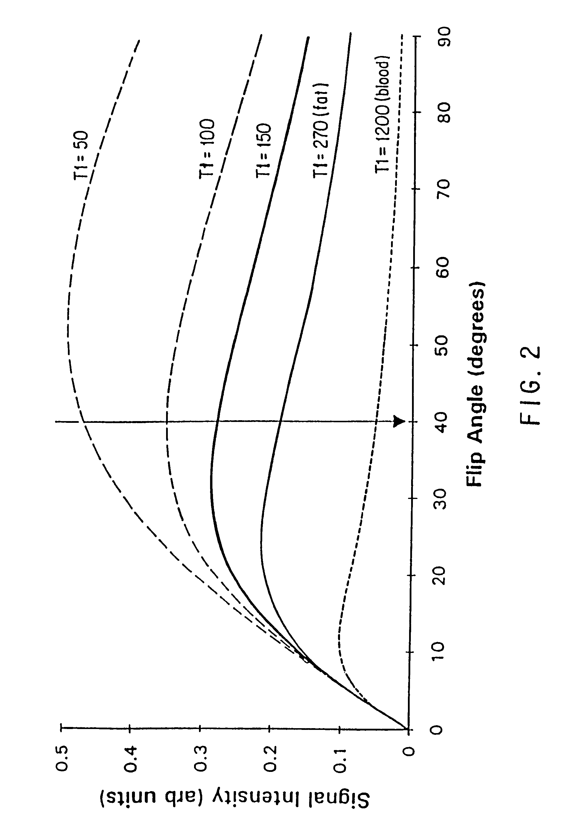
[0172]All imaging was performed on a 1.5 Tesla superconducting magnet (General Electric Medical Systems, Milwaukee, Wis.) using the body coil and version 4.7 software. A 3D FT, coronal, spoiled, gradient echo volume was acquired centered on the mid-abdomen. The imaging parameters included: 12 cm volume with 60 partitions, 2 mm partition thickness, TR of 25 msec, a TE of 6.9 msec, a flip angle of 400, first order flow compensation, 36 centimeters field of view, 256 by 192 matrix. ...

example 2

[0180]In order to determine the optimal timing of contrast administration, two methods of dynamic administration, bolus and continuous infusion, were compared to non-dynamic injections and to conventional time-of-flight imaging.

[0181]Gadolinium enhanced magnetic resonance arteriography was performed in 52 patients referred for routine MRA of the abdominal aorta or branch vessels. Imaging was performed as described in Example 1. The total acquisition time was 5:08 minutes to cover approximately 36 cm of aorta and iliac artery in the superior to inferior dimension. In 20 of the patients of the dynamic gadolinium infusion imaging was performed with 28 partitions each 2 mm thick with a 256 by 256 matrix to reduce the scan time to 3:18 minutes.

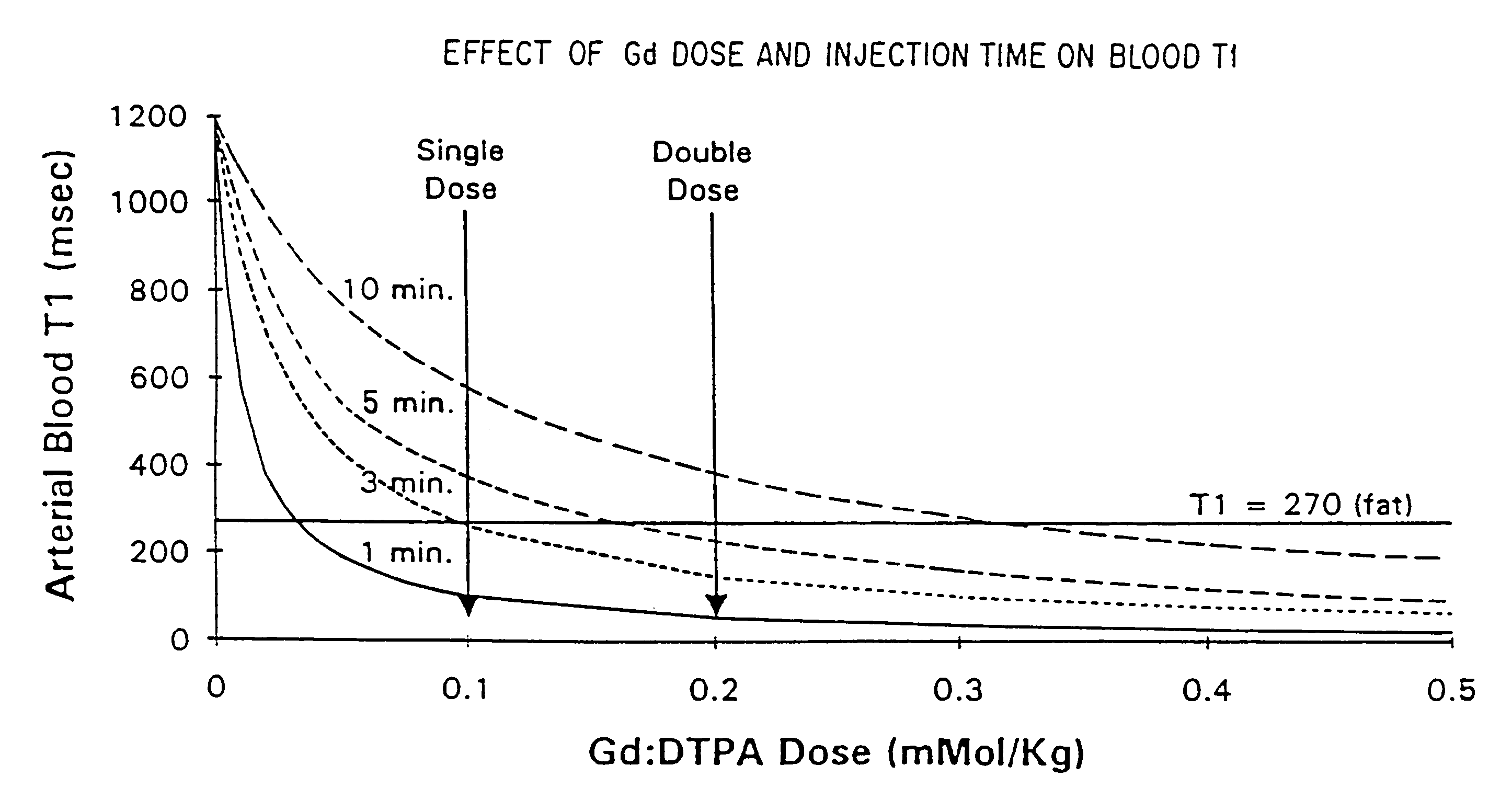
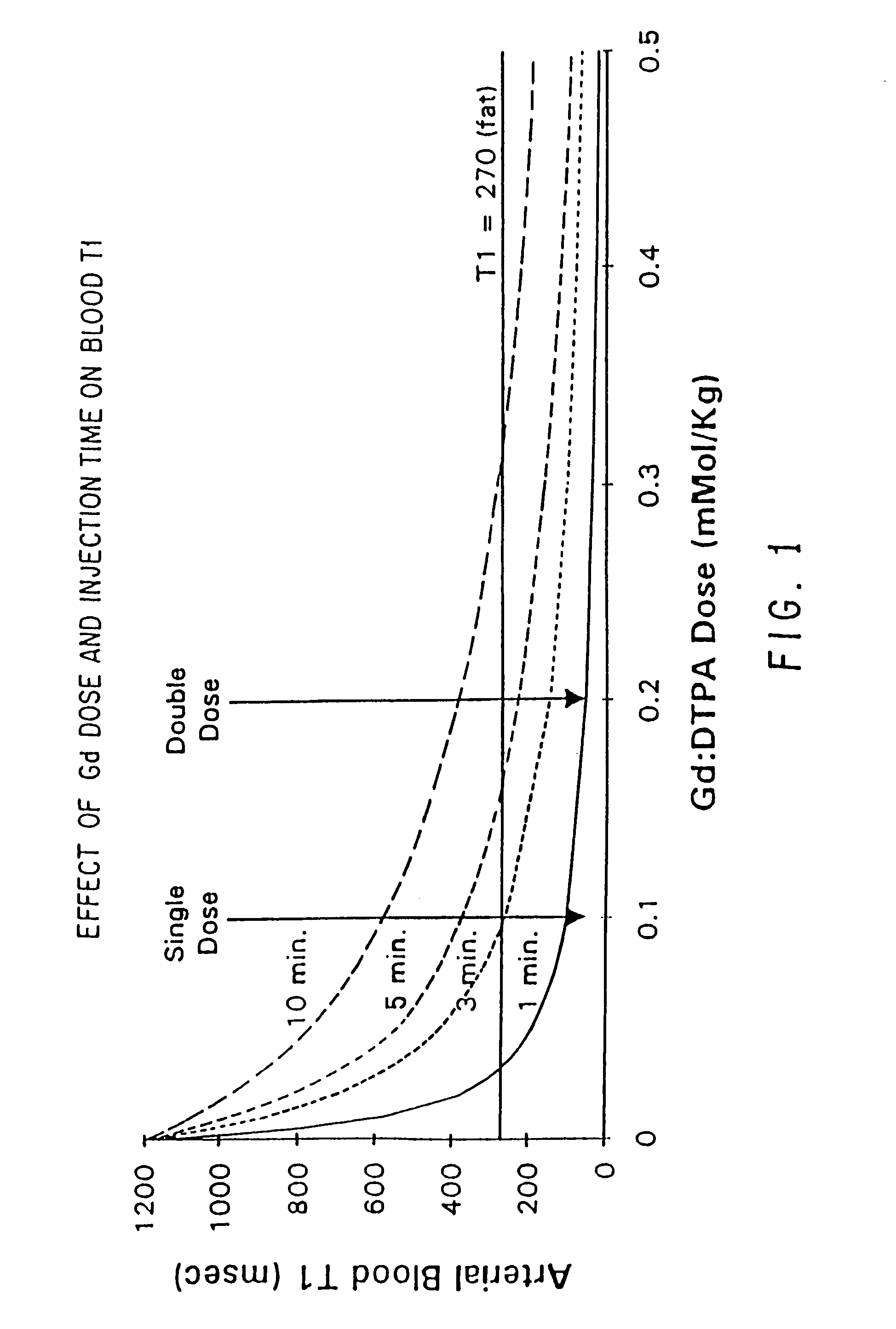
[0182]After pre-scanning, venous access was obtained via a 22 gauge angiocatheter. A dynamic acquisition was then performed during hand injection of gadolinium dimeglumine (Berlex Laboratories, Cedar Knoll, N.J.) 0.2 millimoles / Kg. In 12 patients, ...

example 3

[0191]MRA image data for a patient presenting with an abdominal aortic aneurysm was acquired as described in Example 1. MRA images are shown in FIGS. 10A and 10B.

[0192]The MRA of FIG. 10A depicts the aneurysmal aorta and aneurysmal common iliac arteries as well as severe stenoses of the right external iliac (curved arrow) and inferior mesenteric (straight arrow) arteries and a mild stenosis of the left common iliac artery. The internal iliac arteries are excluded because of their posterior course. FIG. 10B illustrates a digital subtraction angiogram which confirms the findings in FIG. 10A as discussed immediately above.


PUM


Abstract

The present invention is a method and apparatus for providing preferential enhancement of an artery of interest relative to adjacent veins and background tissue. The method and apparatus adapts the timing of a maximum or substantially elevated rate of infusion to correlate with the collection of image data corresponding to the center of k-space. The technique and apparatus temporally correlates the timing of a maximum or substantially elevated rate of infusion and the mapping of k-space according to the location of the artery of interest, the size of the artery of interest, the physical condition of the patient, the time delay due to the configuration of the contrast agent delivery system, and / or the type of pulse sequence employed by the imaging apparatus. Adapting the timing of a maximum or substantially elevated rate of infusion to correlate with the collection of image data corresponding to the center of k-space provides a period of a maximum or substantially elevated contrast concentration in the artery of interest relative to adjacent veins during collection of at least a portion of the image data corresponding to the center of k-space.

Description

[0001]This application is a continuation of application Ser. No. 09 / 899,353, filed Jul. 5, 2001 now U.S. Pat. No. 6,889,072, which is a continuation of application Ser. No. 09 / 061,101, filed Apr. 16, 1998 now U.S. Pat. No. 6,278,892, which is a continuation of application Ser. No. 08 / 705,818, filed Aug. 30, 1993 now U.S. Pat. No. 5,762,065, which is a continuation of Ser. No. 08 / 378,384, filed Jan. 25, 1995 now U.S. Pat. No. 5,553,619, which is a continuation-in-part of application Ser. No. 08 / 071,970, filed Jun. 7, 1993 now U.S. Pat. No. 5,417,213.BACKGROUND OF THE INVENTION[0002]This invention relates to a method of, and apparatus for use in, magnetic resonance angiography to detect, diagnose, and treat arterial diseases and injuries. Arterial diseases and injuries are common and often have severe consequences including death. Imaging arteries serves to detect and characterize arterial disease before these consequences occur.[0003]A conventional method of arterial imaging includes...

Claims


Application Information

Patent Timeline
Patent Type & Authority Patents(United States)
IPC IPC(8): A61B5/05A61K49/18G01R33/28G01R33/561G01R33/563
CPCA61B5/411A61B5/416G01R33/5601G01R33/563A61K49/18A61B5/201A61B5/7285G01R33/56308G01R33/281G01R33/5619
Inventor PRINCE, MARTIN R.
Owner PRINCE MARTIN R
Why Eureka
Try Eureka