Apparatus for detecting and recording cellular responses

a technology of cellular responses and apparatus, applied in the field of apparatus for detecting and recording cellular responses, can solve the problems of less than optimal reproducibility and reliability, additional problems, and difficulty in achieving a highly reproducible and reliable assay or quantitative analysis of electrophysiological responses by conventional manual perfusion and membrane potential measurement techniques

Inactive Publication Date: 2010-09-21
TRUSTEES OF BOSTON UNIV
View PDF6 Cites 0 Cited by
  • Summary
  • Abstract
  • Description
  • Claims
  • Application Information

AI Technical Summary

Benefits of technology

[0015]One embodiment of the invention is directed to cellular physiology workstations that enable automated execution of experimental protocols for electrophysiological experiments and for the development of more complex protocols based on extended recording sessions. As currently developed for oocyte electrophysiology, the apparatus comprises one or more custom-built recording chambers, a perfusion control system designed for rapid application of about 2 to about 16 or more solutions under automated control, software-based virtual instrumentation developed to automate the execution of experimental protocols, and a data acquisition and control platform which integrates the entire system. The system is fully customizable through a sophisticated object-oriented programming language and can be easily adapted to applications such as patch clamp electrophysiology, calcium imaging studies, confocal microscopy and other applications where perfusion control and data acquisition need to be tightly integrated.

Problems solved by technology

It has been difficult to achieve a highly reproducible and reliable assay or to achieve quantitative analysis of electrophysiological response by conventional manual perfusion and membrane potential measurement techniques.
These techniques have many shortcomings because of variabilities due to human errors, operator fatigue and inconsistencies between operators, and less than optimal reproducibility and reliability.
Further, the perfusion and detection steps typically require long and complicated manual manipulations which create additional problems.
The cultured cell becomes less viable with time and it is difficult to control the temperature and oxygen tension.
The limited dexterity of even the most experienced operator limits the number of experiments may be performed on one cell.
Reliance on human operators has resulted in reaction times that are considerably longer than theoretically possible.
These systems have suffered generally from inability to individually measure a physiological response of a cell.
The system is limited to analysis of cell response of a through transmission microscopy.
The system is limited to the monitoring of microorganism growth and multiplication by an impedance measuring process.
Capco's disclosed method is limited to contacting the oocytes to one solution comprising a candidate tumor promoting compound.

Method used


Image

  • Apparatus for detecting and recording cellular responses
  • Apparatus for detecting and recording cellular responses
  • Apparatus for detecting and recording cellular responses

Examples


example 1

Oocyte Isolation

[0111]Female, oocyte positive Xenopus laevis frogs, purchased from Nasco, Inc. were kept on an about 12 hour light / about 12 hour dark cycle. Frogs were maintained on a diet of chopped calf liver fed about every three days. Prior to surgery, frogs were anesthetized in a solution containing about 0.15% Tricaine for about 30 minutes. Ovarian sections were removed through a lateral abdominal incision, after which the incision was sutured with about 4 to about 5 stitches and the animal allowed to recover in isolation for about 3 hours to about 4 hours. Ovarian lobules containing follicular oocytes were immediately placed in calcium-free ND96 solution (96 mM NaCl, 1 mM MgCl2, 2 mM KCl, 50 mM Hepes, 2.5 mM pyruvate) and cut into groups of about 10 to about 20 oocytes. Following a treatment with collagenase (Sigma, type II, 2 mg / ml) at about 2 mg / ml for about 2 hours at room temperature, individual oocytes were obtained free of their follicular layer. Selected Dumont stage V...

example 2

RNA Preparation

[0112]RNA was prepared for injection into oocytes by extraction of mRNA from brain tissue and by synthesis using in vitro transcription of linearized DNA templates encoding recombinant receptor subunits.

[0113]The extraction technique of RNA preparation uses brain mRNA from chick embryos of about 19 day old as starting material. Extraction was performed using the Dynabeads Oligo-dT(25) isolation kit (Dynal, Inc.) which utilizes magnetic beads having an attached poly-thymidine oligomer to allow magnetic separation of poly(A)+ RNA from cell homogenates.

[0114]In vitro transcription was performed using plasmids containing the GluR3 (flop) and GluR6 cDNA as starting material. Plasmids were linearized with restriction endonuclease XhoI (GluR3) or XbaI (GluR6) prior to in vitro transcription with T3 RNA polymerase using a commercially available kit (Message Machine; Ambion, Inc.; Austin, Tex.).

example 3

Electrophysiology

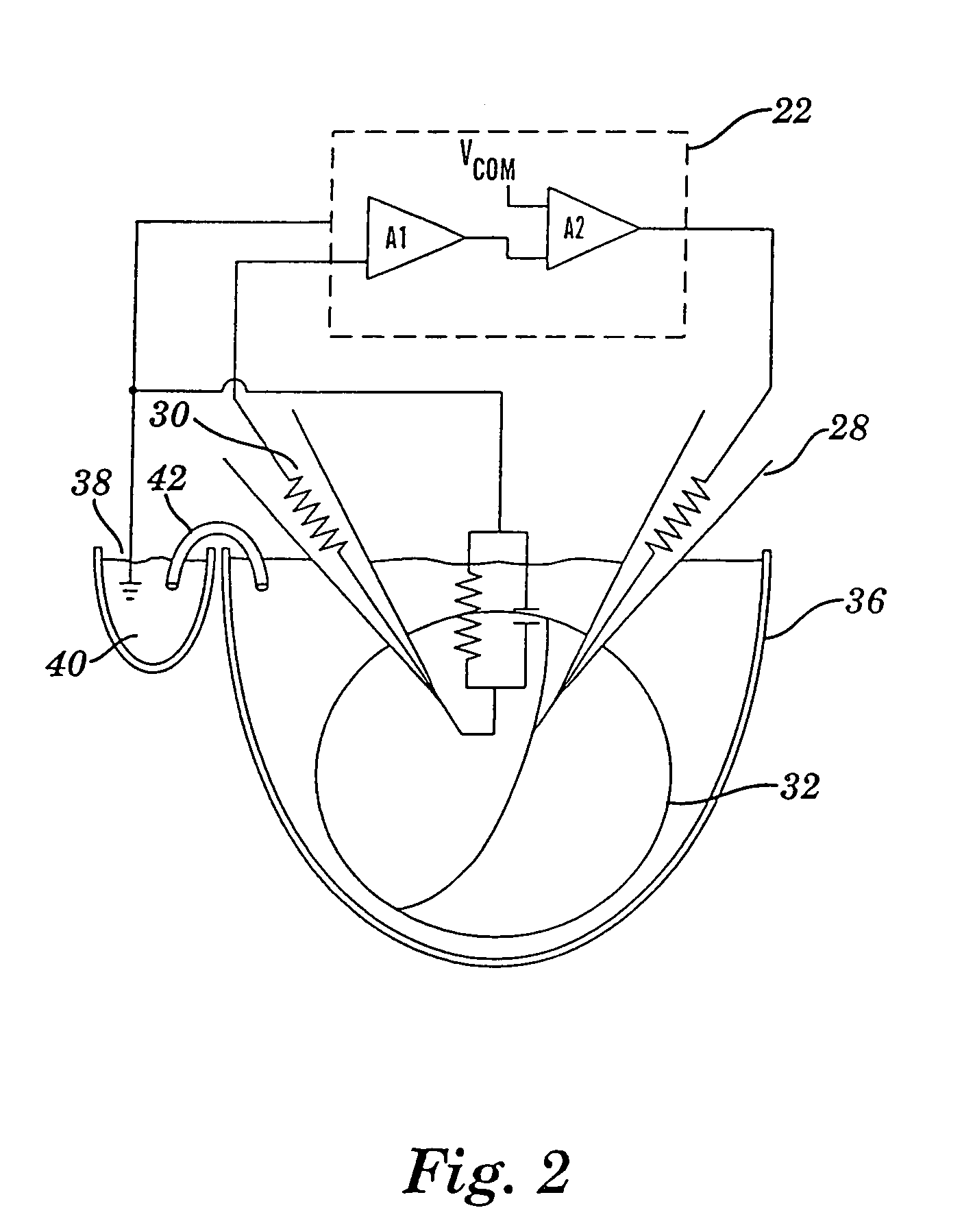
[0115]About 3 days to about 5 days after RNA injection, electrophysiological recordings were carried out using an Axoclamp-2A voltage clamp amplifier (Axon Instruments, Inc.). Experiments were performed in two-electrode voltage clamp mode using two intracellular microelectrodes of about 1 to about 3 mega-ohm resistance filled with a solution of about 3M KCl. A close-up view of an oocyte under impalement in voltage-clamp mode is shown in FIG. 7.

[0116]Oocytes were usually clamped at a holding potential of about −60 mV and stepped to about −100 mV during agent application. GluR6 injected oocytes were treated for about 10 minutes with a solution of about 10 μg / ml concanavalin A to prevent fast desensitization of kainate responses.


PUM

PropertyMeasurementUnit
rise timeaaaaaaaaaa
rise timeaaaaaaaaaa
rise timeaaaaaaaaaa

Abstract

Cellular physiology workstations for automated data acquisition and perfusion control are described. The cellular physiology workstation may be used for physiological and electrophysiological experiments. Methods for employing such cellular physiology workstations in physiological and electrophysiological experiments are also disclosed. The cellular physiology workstations comprise one or more recording chambers each for holding one or more cells to be measured. One or more cells are place in each recording chamber. Perfusions means, such as an automatic perfusion system is connected to the recording chamber to perfuse the cells with a plurality of solutions containing different concentration of one or more agents to be tested. Biosensors, such as patch clamps, electrodes, or microscopes are positioned to detect a response from the cell. The cellular physiology workstation may optionally comprise injecting means for introducing an injection solution into the cell before and during analysis.

Description

CROSS-REFERENCE TO RELATED APPLICATIONS[0001]This application is a continuation application of U.S. patent application Ser. No. 10 / 705,755, filed Nov. 10, 2003; which is a continuation application of U.S. application Ser. No. 09 / 918,370, filed Jul. 30, 2002; which is a continuation-in-part of U.S. application Ser. No. 09 / 519,109, filed Mar. 6, 2000; which is a divisional application of U.S. application Ser. No. 08 / 888,691, filed Jul. 7, 1997; which is a divisional application of PCT / US96 / 18832, filed Nov. 8, 1996; which claims benefit of U.S. Provisional Application No. 60 / 006,326, filed Nov. 8, 1995, the entire content of each of these applications are herein incorporated by reference.GOVERNMENT SUPPORT[0002]This invention was made, in part, with United States Government support under grant number MH-49469, awarded by the National Institute of Mental Health, and the United States Government has certain rights in the invention.MICROFICHE APPENDIX[0003]One microfiche appendix is file...

Claims


Application Information

Patent Timeline
Patent Type & Authority Patents(United States)
IPC IPC(8): C12Q1/02C12M1/36C12M3/00G01N33/487G01N33/50G01N33/68
CPCG01N33/48728G01N33/5008G01N33/502G01N33/5044G01N33/6872
Inventor FARB, DAVID H.YAGHOUBI, NADERGIBBS, TERRELL T.
Owner TRUSTEES OF BOSTON UNIV
Why Eureka
Try Eureka