
Immuno-PCR method for detecting nasopharyngeal carcinoma markers and kit thereof
a nasopharyngeal carcinoma and immunopcr technology, applied in the field of immunopcr method for detecting nasopharyngeal carcinoma and kit, can solve the problems of death of patients, difficult to kill or remove cancer cells completely, all diagnostic tools have their own limitations, etc., and achieve the effect of increasing the signal value and raising the sensitivity
- Summary
- Abstract
- Description
- Claims
- Application Information
AI Technical Summary
Benefits of technology
Problems solved by technology
Method used
Image

Examples
embodiment 1
nd Modification of Substrate Surface
[0057]Because the stability of protein is relatively poor, it is difficult to maintain its biochemical activity for a long time, and it is apt to be influenced by the experiment condition (solvent type, temperature, pH value etc.) and cause the change or modification of protein structure and the loss of activity. Therefore it is necessary to carry on the chemical modification of substrate or add the extra additive, in order to maintain the structure and biochemical activity of protein. According to the characteristics of substrate and immobilized molecules, suitable immobilization method can be selected. Its basic steps are: (a) The activation of substrate: the specific functional group is produced on the substrate surface after chemical modification. The functional group can bind the protein. (b) The immobilization of protein: the protein can be bound to the activated substrate through the functional groups (eg., amino group, carboxyl group) of a...
embodiment 2
on of Markers
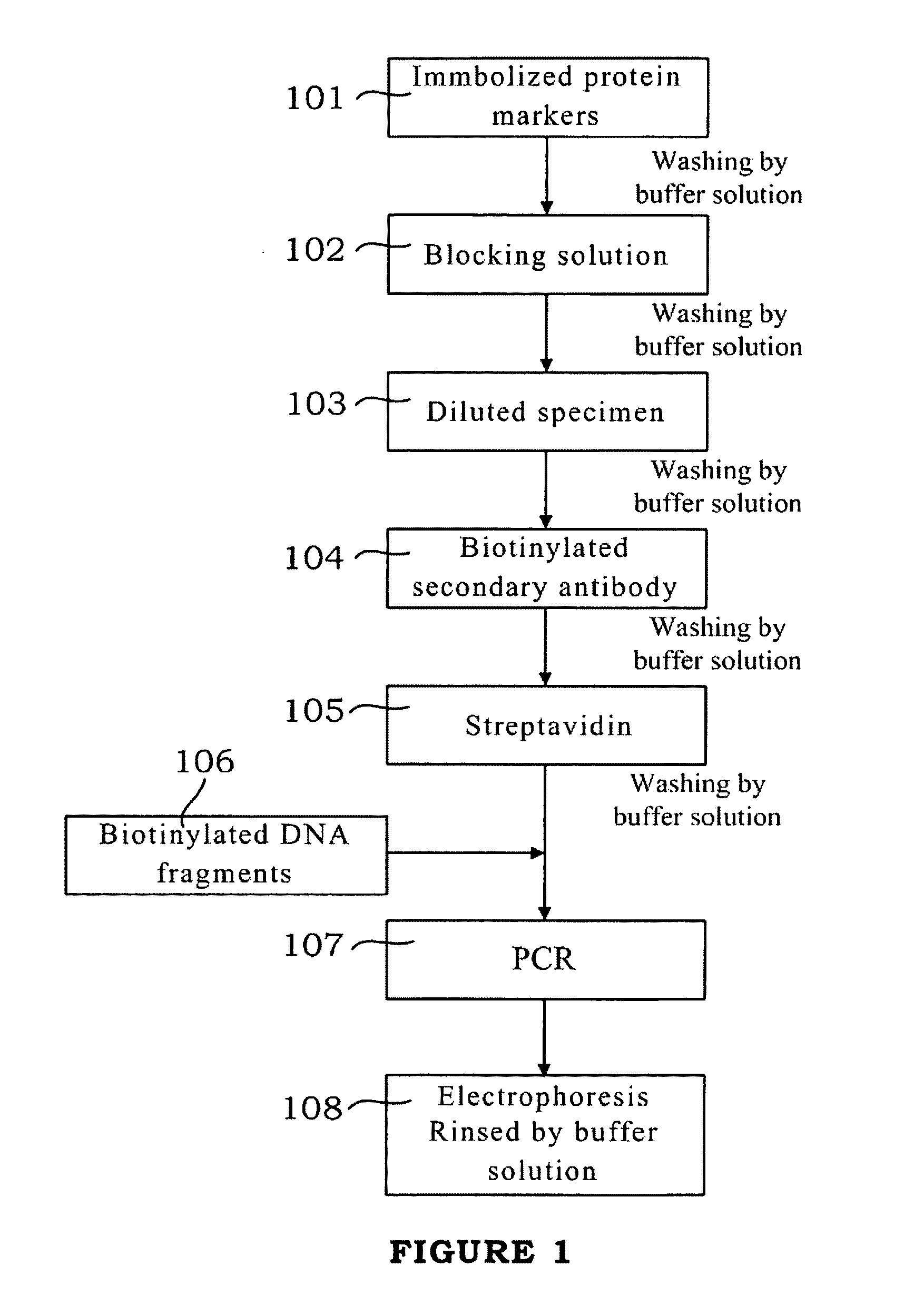
[0063]Please refer to FIG. 1, which is the schematic flow diagram of the present invention. As shown in Step 101, 50 μL of EBNA1 (10 μg / mL) is added on the modified glass substrate, and reacted at 37° C. for 4 hours. The buffer solution (10 mM Tris, pH 7.3, 150 mM NaCl) is used to wash out un-reacted antigens and impurities. Then according to Step 102, the blocking solution (10 mM Tris / HCl, pH 7.6, 6% skimmed milk power, 0.2% NaN3, 0.05% Tween-20 and 5 mM EDTA) is added. After reacting at 37° C. for 1 hour, the glass substrate is rinsed by the buffer solution.
embodiment 3
ng Reaction
[0064]Refer to Step 103 in FIG. 1, the diluted serum is added to be tested. It is reacted at 37° C. for 1 hour. The specific antibody in the serum is combined with the antigen immobilized on the substrate surface to form the solid phase antigen-antibody complex. After it is washed by the buffer solution, only the specific antibody is left on the substrate, and other compositions in the serum will be washed out. Then 50 μl of biotinylated antibody (0.25 μg / ml of concentration) is added, and reacted at 37° C. for 1 hour (Step 104).
[0065]After washing by the buffer solution again, the streptavidin (100 ng / ml) is added and incubated at the room temperature for 30 minutes (as shown in Step 105). As shown in Step 106, after washing, biotinylated DNA fragments (1 ng / ml) is added, and reacted at 37° C. for 1 hour. After it is washed completely, the amount of biotinylated DNA fragments will be proportional to the amount of antibody to be tested in the sample.
PUM
| Property | Measurement | Unit |
|---|---|---|
| pH | aaaaa | aaaaa |
| pH | aaaaa | aaaaa |
| concentration | aaaaa | aaaaa |
Abstract
Description
Claims
Application Information