
Biopolymer system for tissue sealing

a biopolymer and tissue technology, applied in the field of tissue sealants, can solve the problems of dimensional instability of both synthetic hydrogels, nerve or brain tissue pressure can produce serious side effects, and undergo considerable swelling

Active Publication Date: 2010-12-21
ENDOMEDIX
View PDF71 Cites 37 Cited by
  • Summary
  • Abstract
  • Description
  • Claims
  • Application Information

AI Technical Summary

Benefits of technology

[0016]The invention further provides a tissue sealant that due to its exceptional dimensional stability may be used in situations where swelling and the resulting pressure are undesirable and produce unwanted side effects.
[0017]The invention further provides a tissue sealant that offers a very low risk of contamination by pathogens such as viruses and prions.
[0025]The invention further provides methods for using a hydrogel according to the present invention in tissue repair and other medical procedures. A specific embodiment of a hydrogel according to the present invention is used to reinforce a suture line, or to seal cut, torn, or perforated tissues. It is also used to prevent leakage of biological fluids, such as cerebrospinal fluid, through repair of biological membranes that when intact contain the fluids. It is used to bring tissues into approximation and hold them in place after a surgical procedure has been carried out.
[0029]In another specific embodiment, the therapeutic agent may be an antibiotic to inhibit bacterial infection after repair of a wound or after damage to tissues caused by surgery. Or, a protective agent may be an anti-inflammatory substance wherein it is advantageous to supply the substance directly at the site of damage that is repaired with the tissue sealant, such as to reduce swelling and resulting pressure on surrounding tissues.
[0032]Thus, the hydrogels of the present invention, and their uses as tissue sealants, as media further containing therapeutic or protective agents, and as tissue sealants further containing therapeutic or protective agents, offer outstanding advantages of ease of use, biocompatibility and biodegradability, suitability for use in conjunction with other surgical procedures, strength, adhesivity, and versatility.

Problems solved by technology

However, bovine tissues are also a source of concern in terms of the possible presence of pathogenic entities such as viruses or prions.
However, both of these synthetic hydrogels are dimensionally unstable in the presence of water, undergoing considerable swelling.
This tendency to swell can be highly disadvantageous in certain applications, such as neurosurgery, where the resulting pressure on nerve or brain tissue can produce serious side-effects.
The material is known to be non-toxic and biocompatible, and since chitin is not derived from vertebrates and is processed under rather harsh conditions such as exposure to alkalai during its transformation into chitosan, the possibility of contamination with viruses or prions that are pathogenic to mammals is very low.

Method used


Image

  • Biopolymer system for tissue sealing
  • Biopolymer system for tissue sealing
  • Biopolymer system for tissue sealing

Examples


example 1

Preparation of Acrylated Chitosan

[0117]

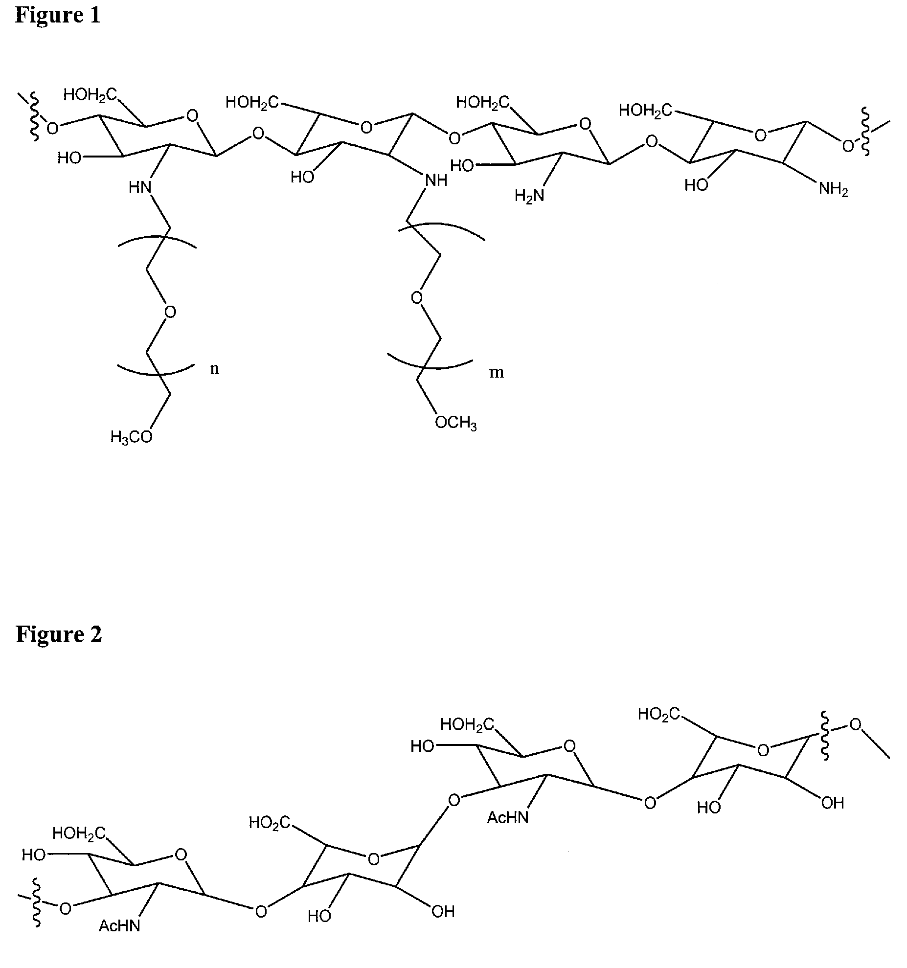
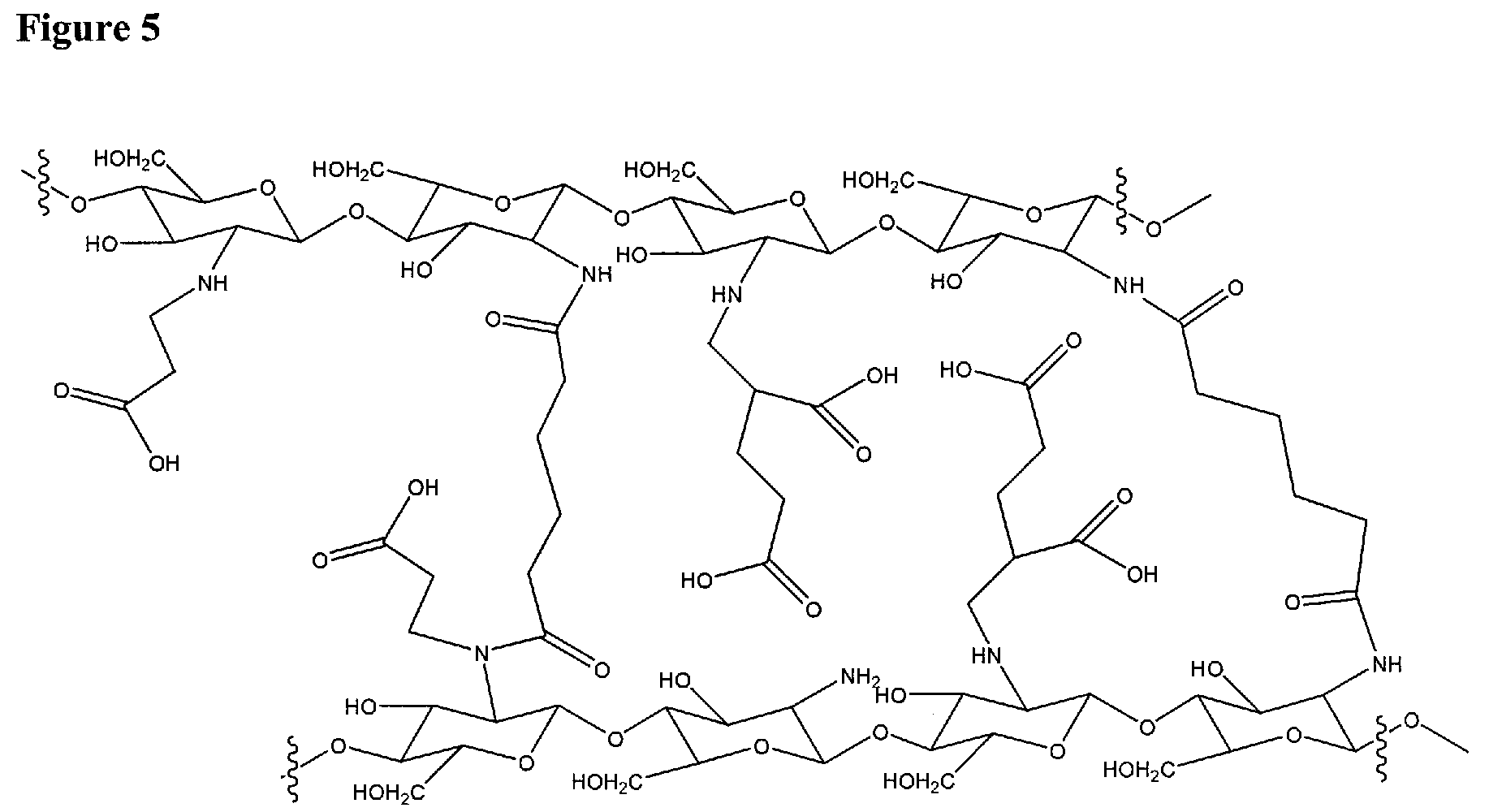
[0118]5.52 ml of acrylic acid was dissolved in 150 ml of double distilled water and 3 g of chitosan (Kraeber® 9012-76-4, molecular weight 200-600 kD) was added to it. The mixture was heated to 50 C and vigorously stirred for 3 days. After removal of insoluble fragments by centrifugation, the product was collected and its pH was adjusted to 11 by adding NaOH solution. The mixture was dialyzed extensively to remove impurities.

example 2

Preparation of PEG-Chitosan

[0119]

[0120]Monomethyl-PEG-aldehyde was prepared by the oxidation of Monomethyl-PEG (MPEG) with DMSO / acetic anhydride: 10 g of the dried MPEG was dissolved in anhydrous DMSO (30 ml) and chloroform (2 ml). Acetic anhydride (5 ml) was introduced into the solution and the mixture is stirred for 9 h at room temperature. The product was precipitated in 500 ml ethyl ether and filtered. Then the product was dissolved in chloroform and re-precipitated in ethyl ether twice and dried.

[0121]Chitosan (0.5 g, 3 mmol as monosaccharide residue containing 2.5 mmol amino groups, Kraeber 9012-76-4, molecular weight 200-600 kD) was dissolved in 2% aqueous acetic acid solution (20 ml) and methanol (10 ml). A 15 ml sample of MPEG-aldehyde (8 g, DC: 0.40) in aqueous solution was added into the chitosan solution and stirred for 1 h at room temperature. Then the pH of chitosan / MPEG-monoaldehyde solution was adjusted to 6.0-6.5 with aqueous 1 M NaOH solution and stirred for 2 h at...

example 3

Preparation of a PEG-Chitosan and Hyaluronan Hydrogel

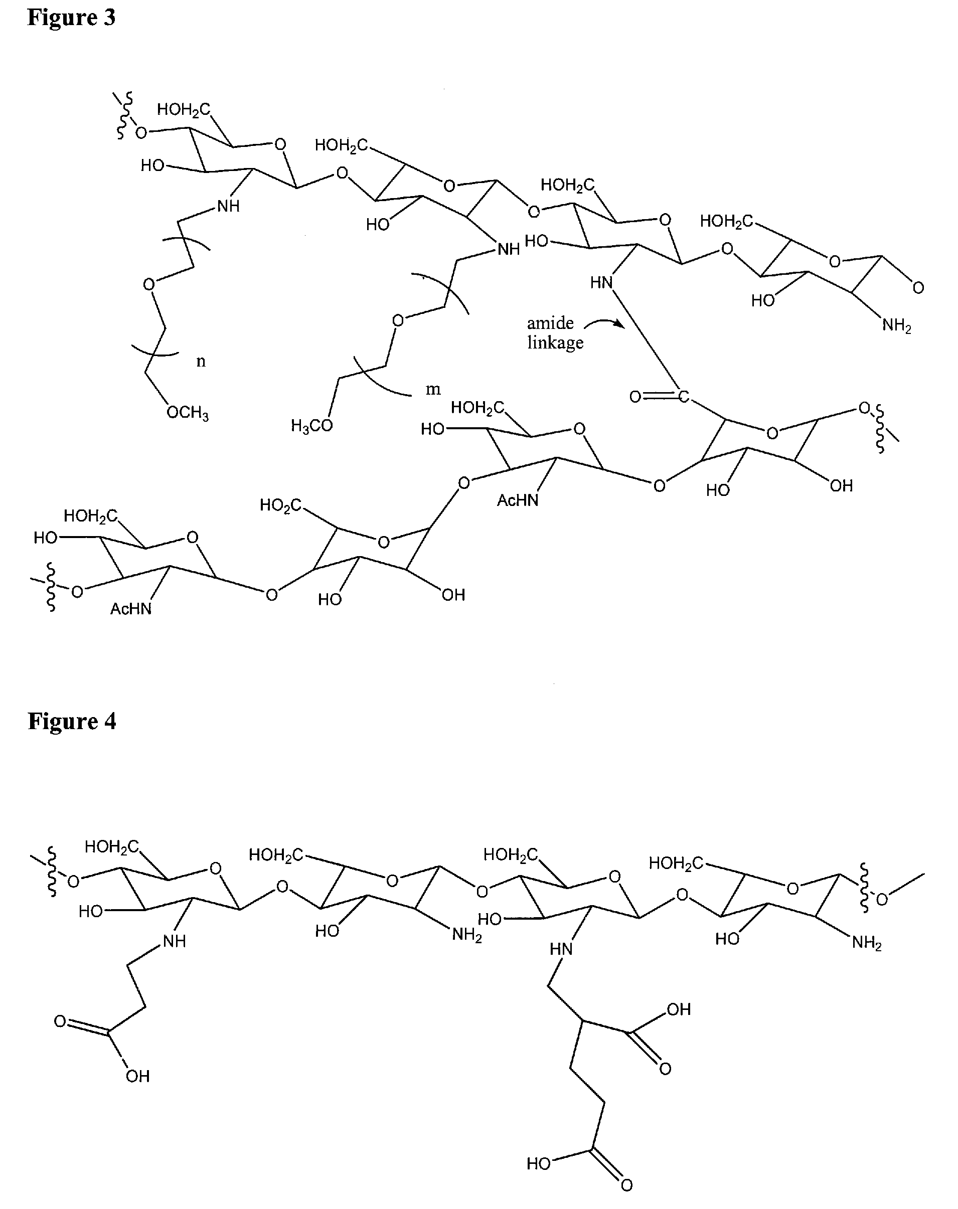
[0122]Hyaluronan (sodium hyaluronate, Kraeber 9067-32-7) was dissolved in water as a 0.5% solution by weight. PEG-chitosan, prepared as described in Example 2, was dissolved in water as a 5% solution by weight. A sample of each solution (0.5 mL of each) was mixed, then a solution of EDCI (20 μL of a solution in water at 350 mg / mL) was added and the solution was thoroughly mixed. Immediately a solution of N-hydroxysuccinimide (20 μL of a solution in water at 125 mg / mL) was added and thoroughly mixed in to form a premix. The premix gelled into a hydrogel in about 7 minutes at ambient temperature (22° C.). At 37° C. gelation occurred in about 2 minutes.


PUM

PropertyMeasurementUnit
body temperatureaaaaaaaaaa
temperatureaaaaaaaaaa
pHaaaaaaaaaa

Abstract

A tissue sealant for use in surgical and medical procedures for sealing the tissues of a living mammal is provided. The tissue sealant comprises a hydrogel which is formed by gelation of a premix disposed on the tissue to be sealed. The premix comprises alkylated chitosan or a gelatin, and a polybasic carboxylic acid or an oxidized polysaccharide, in an aqueous medium. The premix can also include a dehydrating reagent, a carboxyl activating reagent, or both. A specific use of the tissue sealant is in the repair of the dura mater after brain surgery to prevent leakage of cerebrospinal fluid. The tissue sealant may include a therapeutic or protective agent such as an antibiotic or an anti-inflammatory drug.

Description

CLAIM OF PRIORITY FROM A PRIOR-FILED APPLICATION[0001]This application is a continuation-in-part of U.S. patent application Ser. No. 11 / 379,182, filed Apr. 18, 2006, which is incorporated herein by reference, and claims the priority thereof.FIELD OF THE INVENTION[0002]The invention relates to tissue sealants for medical or veterinary use, methods of preparing the sealants, and methods of using them in medical procedures.BACKGROUND OF THE INVENTION[0003]Tissue sealants are increasingly important adjuncts in surgical procedures, being used in fields such as vascular surgery, cardiac surgery, spine surgery and brain surgery as well as in general surgery. Uses for tissue sealants include, among others, augmenting or replacing sutures to join tissues or place them in proximity, closing perforations in biological membranes to prevent leakage of fluids, incorporating medicinal substances at the location of emplacement for localized release, and filling areas of tissue removal. One commonly...

Claims


Application Information

Patent Timeline
Patent Type & Authority Patents(United States)
IPC IPC(8): A61K48/00
CPCA61K31/722A61K38/1875A61K47/48215A61K47/48784A61K48/00A61K49/0002A61K49/0457A61K51/1213A61L24/0031A61L24/043A61L24/08A61L24/104A61K31/721C08L89/00C08L5/08A61K9/0085A61K9/06A61K47/60A61K47/6903A61P17/02A61P19/00A61P25/00A61P9/00A61L26/0052A61L26/0066A61L26/008A61L26/009A61L2300/232A61L2300/236
Inventor CHEN, WEILIAMABRAHAMS, JOHN M.
Owner ENDOMEDIX
Learn More