Electromagnetic treatment of tissues and cells

a tissue and cell technology, applied in the field of biomedical engineering, biochemistry and medical treatment and surgical procedures, can solve the problems of affecting the healing process, and affecting the healing effect of the patient, so as to achieve the effect of convenient application

Inactive Publication Date: 2011-06-28
ROCKY MOUNTAIN BIOSYST
View PDF13 Cites 1328 Cited by
  • Summary
  • Abstract
  • Description
  • Claims
  • Application Information

AI Technical Summary

Benefits of technology

[0087]FIG. 35 depicts fusion composition in the form of a laminate, or tape, which can be easily applied to the tissue to be anastomosed.

Problems solved by technology

Failure to close surgical wounds optimally can also result in serious and excessive scarring.
The healing process may also complicate the status of the patient through formation of scar tissue.
This scarring helps to close the wound, but its formation is accompanied by contraction and buildup of tissue which can lead to a loss in flexibility at the wound site and, in severe cases, may result in loss of mobility to the patient.
While these methods are generally successful, at times complications arise due to inadequate closure of the wound that could result in the tissues separating or in “leakiness.” In particular, the quality of suturing depends on manual dexterity of the surgeon and adequate access to the wound.
Current designs of surgical clips can slip if applied incorrectly or accidentally disturbed.
Surgical clips can also cause damage to the vessels or structures to which they are applied if the surgeon applies excessive compression force.
With the increasing use of minimally invasive surgical methods, such as endoscopy, wound access and the efficient closure of wounds has become a significant issue in medicine.
Nonetheless, conventional stapling is limited in that it usually requires an anvil be placed behind the tissues to be joined, and that enough space is available to produce the necessary force to form the staple against the anvil.
Sutures cannot easily be secured by traditional methods through an endoscope and current stapling methods generally require an anvil be placed behind the tissue thereby limiting their use.
Nevertheless, these devices are generally unsuitable for the purpose of occluding a wound thereby enhancing long-term healing.
Use of lasers for tissue welding appeared very promising, however, over the years the techniques have been shown to present certain limitations.
The laser energy must be manually directed by the surgeon which leads to operator variability.
Additionally, the radiant energy is not dispersed evenly throughout the tissue.
The high energy at the focal point may result in local burns and the heating effect drops off rapidly at a small distance from the focal point.
Finally, lasers are expensive and currently cannot be miniaturized easily.
Nevertheless, the lack of control and the inability to induce uniform heating in the nerve as a result of laser irradiation restricts the utility of laser-nerve welding to the laboratory.
Use of lasers for tissue welding appeared very promising, however, the techniques have certain limitations.
The laser energy must be manually directed by the surgeon, which leads to operator variability.
Additionally, the radiant energy is not dispersed evenly through the tissue.
The high energy at the focal point may result in local burns and the heating effect drops off rapidly at a small distance from the focal point.
Finally, lasers are expensive and currently cannot be easily miniaturized.
The prior art fails to describe adequate means for delivering compositions that may be activated using radiofrequency energy sources.
Common problems exist throughout the prior art.
These include, for example, tissue damage due to uneven heating, unknown and / or uncontrollable thermal history, i.e., time-temperature profile, and relatively high cost.
The Code of Federal Regulations, 21 CFR 860.7(e)(1), establishes that there is “reasonable assurance that a device is effective when it can be determined, based upon valid scientific evidence, that in a significant portion of the target population, the use of the device will provide clinically significant results.” Devices that cannot be shown to provide consistent results between patients, or even within a patient upon multiple use, will have minimal utility and may not be approvable for broad use.
The literature is deficient in descriptions whereby biomolecules are heated through induction.
Treatment protocols for these complications increase morbidity, mortality, hospital time and expense.
Manual compression has been the standard of care for closure after percutaneous coronary interventions, but it requires prolonged bed rest, e.g. 4-12 hours, leading to delayed ambulation, significant medical staff time and associated higher costs.
The routine administration of anticoagulant medication to prevent blood clots and stroke during the diagnostic or interventional procedure can further delay sealing the vessel and postpone ambulation.
Reported complications include an increase in the number of access site infections, as well as pain and discomfort for the patient.
Data on complications is mixed, with several studies showing minor complications comparable to compression, but an increase in major complications that require surgical repair.
Other studies show an increase in minor complications.
Collagen-based devices seal the vessel, but fail to seal the tract.
Limitations to manual pressure include: (1) the 10 to 20 minutes it typically takes to stop bleeding, occasionally taking up to an hour for difficult cases; (2) patients routinely receiving anticoagulant agents during their treatment thus lengthening the time required to stop the bleeding and leave the clinic; (3) applying too little pressure doesn't stop the bleeding, resulting in excess blood loss; (4) applying too much pressure causing the access to thrombose which requires additional interventions; and (5) manual pressure is labor intensive for the dialysis staff when patients are unable to hold their own site following needle removal.
One challenge associated with successful hemodialysis is vascular access, the method used to access a patient's blood supply.
The complications lead to loss of vascular access and the need for corrective surgery in the vast majority of patients twice per year.
The prior art is particularly deficient in devices and methods for minimally-invasive methods that use electromagnetic energy to controllably alter a biocompatible structure thereby making it adhere to tissue through molecular alterations and / or mechanical shrinkage.

Method used


Image

  • Electromagnetic treatment of tissues and cells
  • Electromagnetic treatment of tissues and cells
  • Electromagnetic treatment of tissues and cells

Examples


example 1

Heating of Test Metal

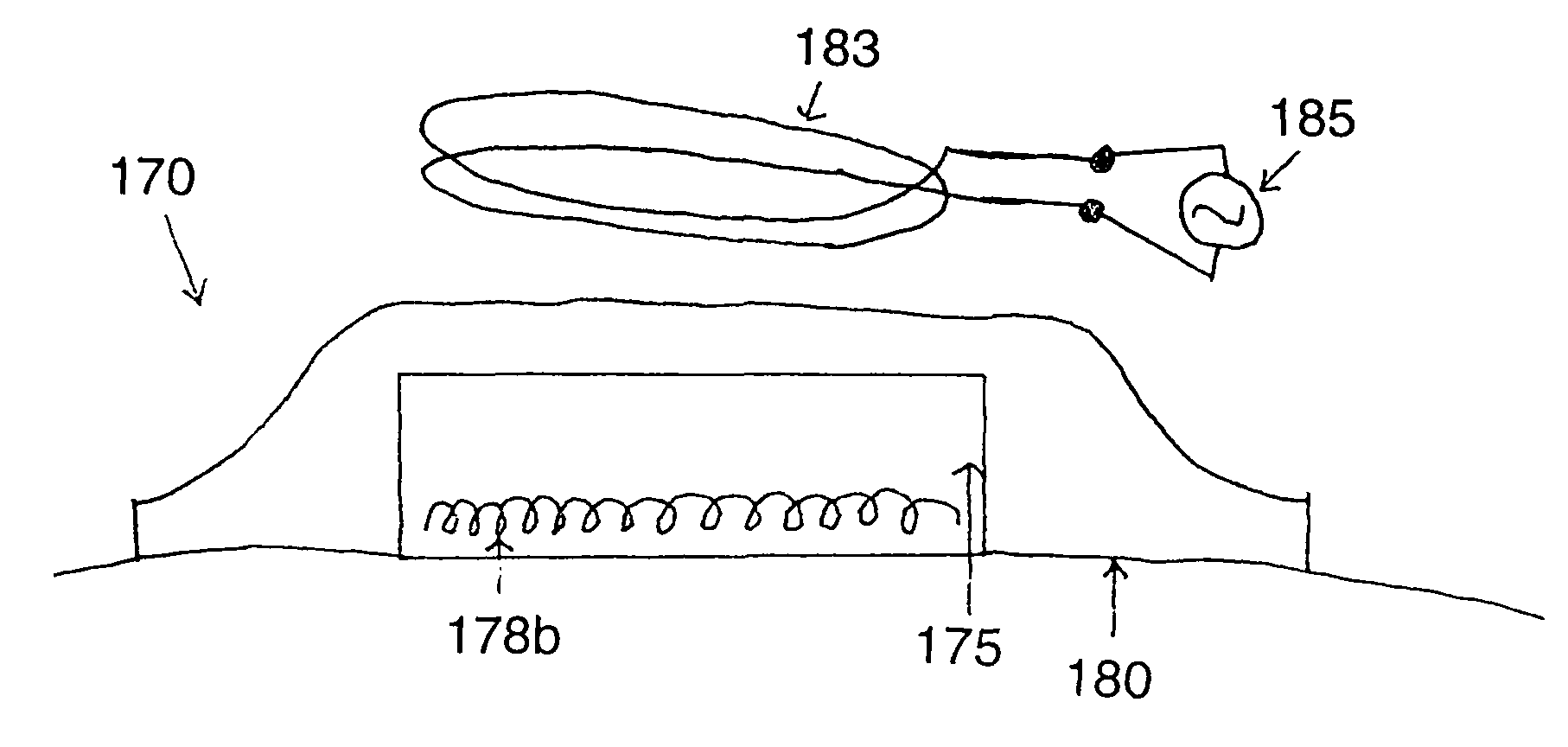
[0317]The prototype activator device was constructed (Quest Product Development, Evergeen, Colo.) and operates at a frequency of about 650 kHz with an output of approximately 210 W. At or near this frequency, the skin depth in tissue, e.g., canine skeletal muscle at 1 MHz, (Francis Duck. Physical Properties of Tissue—A comprehensive reference book. Academic Press: NY, 1990) is about 205 cm and for nickel it is about 14 microns. Thus, no significant heating of tissue occurs as a direct result of the field. Heating only occurs in close proximity to the fusion composition.

[0318]Two applicator designs were used and comprised 200 turns of solid copper wire, 32 and 22 G, resulting in a coil approximately 2.86 cm in diameter and 0.95 cm in width. The bore of the coil was about 0.5 cm. The coils were encapsulated in a Pyrex sleeve through which low-viscosity mineral oil (Sigma-Aldrich Inc., St. Louis, Mo.) is circulated as a coolant. In each of these coils, the magnetic...

example 2

Heating and Coagulating of Test Fusion Formulation

[0321]Fusion formulations were made of 50-75% (w / v) bovine serum albumin or ovalbumin (Sigma-Aldrich, St. Louis, Mo.) in saline with a metal additive of 5% or 10% (w / v) nickel flake with average particle size of about 50 micron (Alfa Aesar, Ward Hill, Mass.) or 10% iron filings with particle size <30 microns (Edmund Scientific, Tonawanda, N.Y.). Approximately 1 ml aliquots of the fusion composition were positioned in thin-walled glass tubes with a diameter of about 4 mm. The tube was positioned in the bore of the applicator. The device was energized for a period of 20-30 seconds. Evidence of denaturation and coagulation was ascertained visually, as the material changed color. This was confirmed by probing the composition with a needle which demonstrated evidence of increased viscosity or stiffness. The composition coagulated with all combinations of applicator and composition. Compositions with more metal or iron versus nickel heated...

example 3

Protein Denaturation

[0322]A radiofrequency electromagnetic device, operating at 650 kHz, was constructed. Near this frequency, the skin depth in tissue, using conductivity values for canine skeletal muscle at 1 MHz, is about 205 cm, while for nickel, it is 14 μm. Two solenoid type coils were constructed using 20 G solid copper wire. The coils were encapsulated in a Pyrex sleeve through which low-viscosity mineral oil is circulated as a coolant. Two coils had 200 turns of solid copper wire, formed into a solenoid, with a diameter of 2.86 cm and width of 0.95 cm. The magnetic intensity within the bore of the coil was calculated to be greater than 100 kA / m, while at approximately 0.5 cm from a single coil face the intensity is calculated to be maximally 0.15 kA / m. Two coils were electronically connected to the radiofrequency power supply and physically arranged with the bore axis parallel and opposing each other with a gap of about 2 cm between the faces of the coils.

[0323]The reactant...


PUM

PropertyMeasurementUnit
frequencyaaaaaaaaaa
skin depthaaaaaaaaaa

Abstract

Provided herein are methods, devices and compositions to conductively or to inductively fix substrates, including tissues, using electromagnetic energy. Also provided is a method of controlling the fixing process via feedback monitoring of a property of the composition and/or of the electromagnetic energy used.

Description

CROSS-REFERENCE TO RELATED APPLICATIONS[0001]This application is a continuation-in-part of U.S. Ser. No. 10 / 736,133, filed Dec. 15, 2003, now U.S. Pat. No. 7,446,803 which is a continuation-in-part of non-provisional U.S. Ser. No. 10 / 441,341, filed May 20, 2003, which claims benefit of provisional U.S. Ser. No. 60 / 381,948, filed May 20, 2002, now abandoned.BACKGROUND OF THE INVENTION[0002]1. Field of the Invention[0003]The present invention relates generally to the fields of biomedical engineering, biochemistry and medical treatment and surgical procedures. More specifically, the present invention provides methods, devices and compositions for inducing changes in biomolecules and bioactive molecules useful for accelerating or enabling certain reactions, fixing or fusing tissues and implants, dressing, sealing or closing a wound to reduce the loss of internal fluids, for enhancing wound healing and for delivery of active agents to tissues.[0004]2. Description of the Related Art[0005]...

Claims


Application Information

Patent Timeline
Patent Type & Authority Patents(United States)
IPC IPC(8): A61B17/02A61B17/00A61B17/04A61B17/064A61B17/068A61B17/11A61B17/115A61B18/04A61B18/08A61B18/14A61F7/00A61F7/12A61L17/00A61L27/50A61N1/40A61N2/02
CPCA61B17/0057A61N2/02A61B17/0487A61B17/064A61B17/068A61B17/1114A61B17/115A61B18/04A61B18/14A61L27/50A61B17/0401A61B18/082A61B2017/0412A61B2017/0427A61B2017/0443A61B2017/0456A61B2017/0462A61B2017/0464A61B2017/0488A61B2017/0641A61B2017/0649A61B2018/00619A61N1/40
Inventor FLOCK, STEPHEN T.MARCHITTO, KEVIN S.
Owner ROCKY MOUNTAIN BIOSYST
Why Eureka
Try Eureka