Methods and devices for microfluidic point-of-care immunoassays
a technology of immunoassays and microfluidic devices, applied in the direction of positive displacement liquid engines, mixers, shaking/oscillation/vibration mixers, etc., can solve the problems of large hybridization chambers, inconvenient or complicated equipment not readily adaptable, and the incubation time is reduced. , the effect of reducing the mean flow velocity
- Summary
- Abstract
- Description
- Claims
- Application Information
AI Technical Summary
Benefits of technology
Problems solved by technology
Method used
Image
Examples
example 1
Preparation and Assembly of a Microfluidic Assay Device
[0140]A microfluidic device for ELISA immunoassay was prepared as follows:[0141]1. Human IgG (0.5 ug in bicarbonate binding buffer pH 9) was deposited onto a plasma-treated test field of a PET sheet. Human IgM was applied as a negative control to a second test field. The proteins were then fixed to the plastic by drying for 5 minutes at 60° C.[0142]2. After fixation, the test fields were blocked for 30 minutes with casein buffer (biotin free) and washed twice with PBST (phosphate buffered saline with 0.1% TWEEN 20).[0143]3. After drying, the treated PET sheets were then assembled between adhesive layers in the microfluidic device of FIG. 3 with integrated pneumatic circuitry for pressurizing the bellows pumps and opening and closing sanitary valves.
example 2
Performance of an ELISA Immunoassay in a Microfluidic Device
[0144]A standard assay for ELISA, useful as a benchmark in method development, involves detection of immobilized human IgG on a solid substrate, followed by blocking and detection of the IgG with biotin-labelled anti-human antibody. The biotin in turn is detected with enzyme-labelled streptavidin.
[0145]Procedure:[0146]1. Referring to the microfluidic device prepared as described in Example 1, anti-human IgG biotin (Pierce, 180 uL, 1:10,000 in casein buffer) was added through the sample port, and the device was incubated for 1 min with slow mixing. The test fields were then rinsed with PBST, with slow mixing for 1 min before removing the rinse. A Microflow microfluidics assay instrument (Micronics, Redmond Wash. USA) was used to perform mixing and washing.[0147]2. Detection reagent (Poly SA-HRP) was added with incubation for 1 min with slow mixing. Poly SA-HRP is streptavidin labeled with horseradish peroxidase. The test fie...
example 3
Assembly of a Microfluidic Device for Immunoassay of Antibodies to Capsular Polysaccharides of Streptococcus pyogenes.
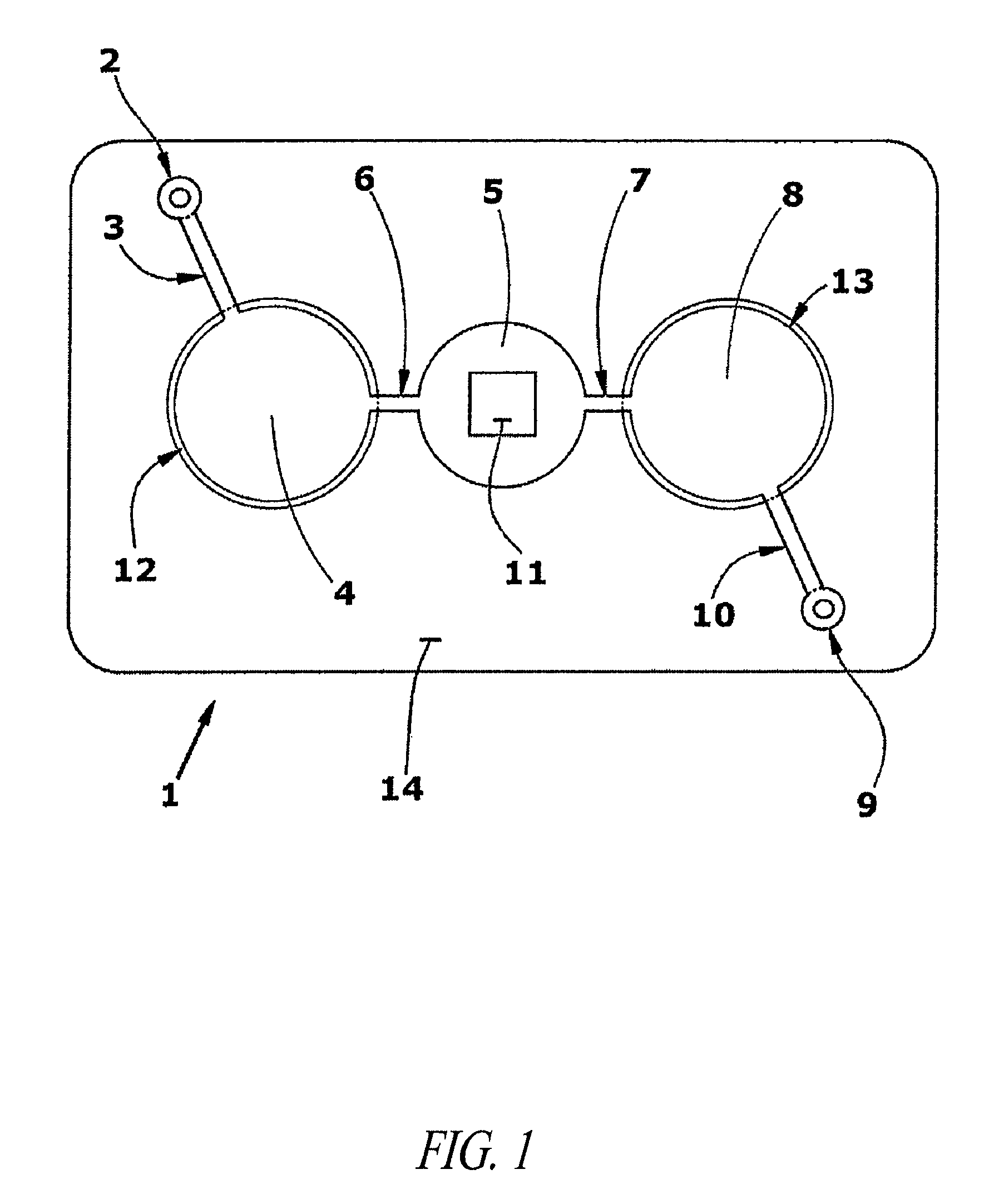
[0151]Assembly of test card:[0152]1. Capsular mucosaccharide antigen purified from Group A Streptococcus pyogenes is immobilized on a N2 plasma activated polyester sheet by co-polymerization with acrylamide monomer. The sheet is previously masked to define the test field area.[0153]2. The plasma treated test field and surrounding areas are then blocked for 30 minutes with casein buffer and washed twice with PBST (phosphate buffered saline with 0.1% TWEEN 20).[0154]3. After drying, a stack of laminate layers including the antigen-treated polyester sheet are assembled in a microfluidic device generally dimensioned as described in FIG. 9 and assembled with fluidic circuitry per the pattern of FIG. 1.
PUM
| Property | Measurement | Unit |
|---|---|---|
| height | aaaaa | aaaaa |
| volumes | aaaaa | aaaaa |
| volumes | aaaaa | aaaaa |
Abstract
Description
Claims
Application Information
 - R&D
- Intellectual Property
- Life Sciences
- Materials
- Tech Scout
- Unparalleled Data Quality
- Higher Quality Content
- 60% Fewer Hallucinations
Browse by: Latest US Patents, China's latest patents, Technical Efficacy Thesaurus, Application Domain, Technology Topic, Popular Technical Reports.
© 2025 PatSnap. All rights reserved.Legal|Privacy policy|Modern Slavery Act Transparency Statement|Sitemap|About US| Contact US: help@patsnap.com


