Stem cell therapy for the treatment of diabetic retinopathy and diabetic optic neuropathy

a technology of optic nerve and stem cell therapy, which is applied in the field of stem cell therapy for the treatment of diabetic optic nerve and diabetic retinopathy, can solve the problems of progressive deterioration of vision, loss of photoreceptors, and abnormal blood vessel formation

Active Publication Date: 2012-11-27
STEMEDICA CELL TECH
View PDF13 Cites 3 Cited by
  • Summary
  • Abstract
  • Description
  • Claims
  • Application Information

AI Technical Summary

Problems solved by technology

This results in loss of photoreceptors through apoptosis or secondary degeneration, which leads to progressive deterioration of vision and, in some instances, to blindness (for a review, see, e.g., Lund, R. D. et al.
This trauma causes the retina to secrete vascularization signals which result in new, abnormal blood vessels being formed.
The thin fragile walls of the abnormal vessels eventually leak blood into the vitreous gel causing vision loss and ultimately blindness.
In the later phases of the disease, continued abnormal vessel growth and scar tissue may lead to further retinal degeneration including retinal detachment and glaucoma.
This is because these procedures only treat abnormal blood vessel formation as it occurs.
They do not offer a lasting solution that addresses the cause of abnormal blood vessel formation which is compromised retinal circulation.
Panretinal photocoagulation also has the undesirable side effect of causing the loss of peripheral vision and compromised night vision.
A drawback of vitrectomy is that it requires a long recovery period during which the patient must remain facing the ground.

Method used


Image

  • Stem cell therapy for the treatment of diabetic retinopathy and diabetic optic neuropathy
  • Stem cell therapy for the treatment of diabetic retinopathy and diabetic optic neuropathy
  • Stem cell therapy for the treatment of diabetic retinopathy and diabetic optic neuropathy

Examples


example 1

Preparation of Multipotent Mesenchymal Stromal Cells (MMSC) from Human Bone Marrow

1. Characteristics and Transport of the Donor Material

[0074]The source for the preparation of human MMSC was a bone marrow suspension (BMS) obtained by puncture of the iliac crest.

[0075]The mandatory clinical, laboratory, and instrumental examinations of the patient (for autotransplantation) were performed, including:

[0076]1. The filling out of the medical history with an attachment of copies of all discharges from the medical history during previous stages of treatment and examination

[0077]2. Complete clinical blood tests

[0078]3. Complete blood biochemistry panel, with determination of renin, aldosterone, and brain natriuretic peptide

[0079]4. Blood group, Rhesus factor

[0080]5. Blood test for HIV and Wasserman test

[0081]6. Blood test for hepatitis B and C markers

[0082]7. Complete immune status

[0083]8. Chest x-ray

[0084]9. Ultrasound of abdominal organs, kidneys

[0085]10. ECG, Halter monitoring

[0086]11. E...

example 2

Immunophenotyping MMSCs

[0153]Probe Preparation. Cells suspended in PBS at concentration 1×10³ cells / ml, fixed in 1% of methanol at 4° C. for 10 min, after this they were washed. Non-specific binding was blocked by incubation in 1% BSA and 0.1% goat serum for 1 hour at room temperature. After this, cells were washed in triple volume of PBS, centrifuged and the pellet suspended in 0.5% primary antibodies solution.

[0154]After incubation, cells were washed in PBS for 40 min. The mouse antibodies used were manufactured by Santa Cruz Biotechnology and Chemicon. As a negative control, nonspecific mouse antibodies were used. The incubation with anti-mouse antibodies, marked with FITC and PE was performed for 20 min. After this, cells were washed in PBS in volume of 1 ml and analyzed using FACS Caliber (BD Biosciences).

Results

[0155]Marker Positive cells (%)

[0156]HLA-ABS-24.9

[0157]HLA-DR 0.7

[0158]CD34-0.29

[0159]CD45-0.76

[0160]CD44-78.4

[0161]CD90-85.7

[0162]CD105-60.5

[0163]Vimentin-96.6

[0164]F(...

example 3

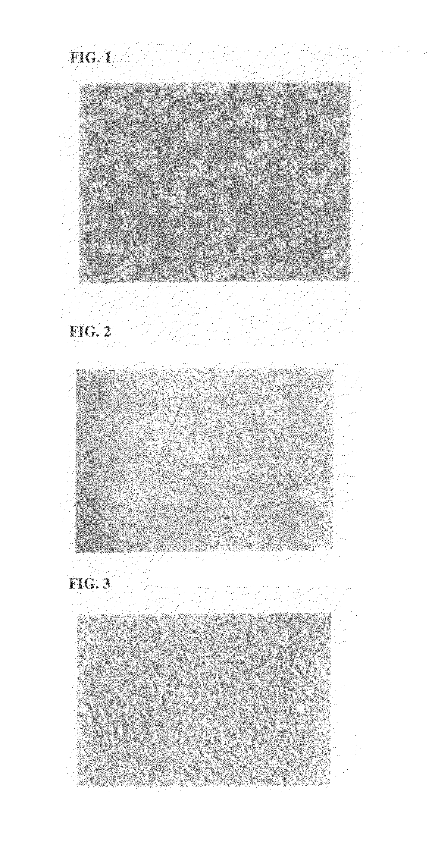
Functional Description of MMSC

[0165]The functional activity of a MMSC culture was evaluated through the ability of the cells to differentiate into mesodermal cell lines (adipose geneses, osteogeneses, chondrogeneses, and miogeneses), under standard conditions, described for differentiating of mesenhymal stem cells, derived from bone marrow (DiGirolamo C. M., Stokes D., Colter D. C. et al. Br. J. Hematol. 1999, 107, 275-281; Sekiya I., Larson B., Smith J. et al. Stem Cells, 2002, 20, 530-541). FIG. 1 presents results obtained for the differentiation experiments.


PUM

PropertyMeasurementUnit
temperatureaaaaaaaaaa
diameteraaaaaaaaaa
volumeaaaaaaaaaa

Abstract

The invention comprises methods and stem cell compositions for the treatment of diabetic retinopathy and other degenerative diseases of the eye. The invention is practiced in two stages with the first stage comprising the administration of neural stem cells to the eye, and the second stage comprising the administration of mesenchymal cells intravenously.

Description

FIELD OF THE INVENTION[0001]The present invention relates to cell-based therapy for ocular diseases and disorders. In particular, the invention provides pharmaceutical compositions, devices and methods for the regeneration or repair of cells and tissues of the retina using mesenchymal and ectodermal stem cells. More particularly, the invention concerns the treatment of diabetic retinopathy and diabetic optic neuropathy through the retinal administration of neural stem cells, and the intravenous administration of mesenchymal cells.BACKGROUND OF THE INVENTION[0002]The patents and publications referred to throughout the specification are incorporated herein by reference as if set forth verbatim.[0003]The retina contains seven layers of alternating cells and processes that convert a light signal into a neural signal. The retinal photoreceptors and adjacent retinal pigment epithelium (RPE) form a functional unit that, in many disorders, becomes unbalanced due to genetic mutations or envi...

Claims


Application Information

Patent Timeline
Patent Type & Authority Patents(United States)
IPC IPC(8): C12N5/07C12N5/073C12N5/074C12N5/077C12N5/0789C12N5/079C12N5/0797A61K35/28
CPCA61K35/28A61P27/00
Inventor GAVRILOVA, NATALIESABURINA, IRINAMIRONOV, NIKOLAY
Owner STEMEDICA CELL TECH
Why Eureka
Try Eureka