Application of electrical stimulation for functional tissue engineering in vitro and in vivo

a technology of functional tissue engineering and electrical stimulation, which is applied in the direction of skeletal/connective tissue cells, prosthesis, therapy, etc., can solve the problem of biocompatibility of the substrate used in the preparative methods, and achieve the effect of enhancing the integration of engineered and host tissues

Inactive Publication Date: 2013-02-05
MASSACHUSETTS INST OF TECH
View PDF12 Cites 13 Cited by
  • Summary
  • Abstract
  • Description
  • Claims
  • Application Information

AI Technical Summary

Benefits of technology

[0012]The present invention is directed to improved systems for the development of bioartificial tissues in vitro and their integration with host tissues following implantation in vivo. More specifically, the present invention provides strategies that (a) allow the formation of three-dimensional tissue-engineered constructs having structural and functional characteristics of a native tissue, and that (b) enhance the integration of engineered and host tissues following implantation of the construct in vivo. The invention further provides three-dimensional tissue-engineered constructs that can be used as bioartificial tissue equivalents for in vitro research and / or as medical implants for the repair and replacement of deficient or damaged natural structure(s).
[0027]In another aspect, the present invention provides methods for treating an individual suffering from tissue deficiency, damage or loss. These methods include producing a three-dimensional tissue-engineered construct according to the preparation methods described above; implanting the construct into the individual in need thereof; and submitting the implanted construct to a biomimetic electrical stimulation. The electrical stimulation may enhance integration of the implanted construct with host tissue, and / or may increase cell survival and / or cell functionality in vivo. The construct to be implanted may further comprise at least one biologically active agent, and thereby serve as vehicle for delivery of the biologically active agent(s) to the individual. The inventive methods of treatment are particularly useful when the tissue to be replaced or supplemented is one that is subject to electrical stimulation in vivo. The inventive methods may, for example, be applied for treating an individual suffering from tissue deficiency, damage or loss associated with a congenital heart disease or with an acquired heart disease.

Problems solved by technology

In some embodiments, the substrate used in the preparative methods is biocompatible.

Method used


Image

  • Application of electrical stimulation for functional tissue engineering in vitro and in vivo
  • Application of electrical stimulation for functional tissue engineering in vitro and in vivo
  • Application of electrical stimulation for functional tissue engineering in vitro and in vivo

Examples


example 1

Preparation of Bioartiflcial Cardiac Muscle Tissue

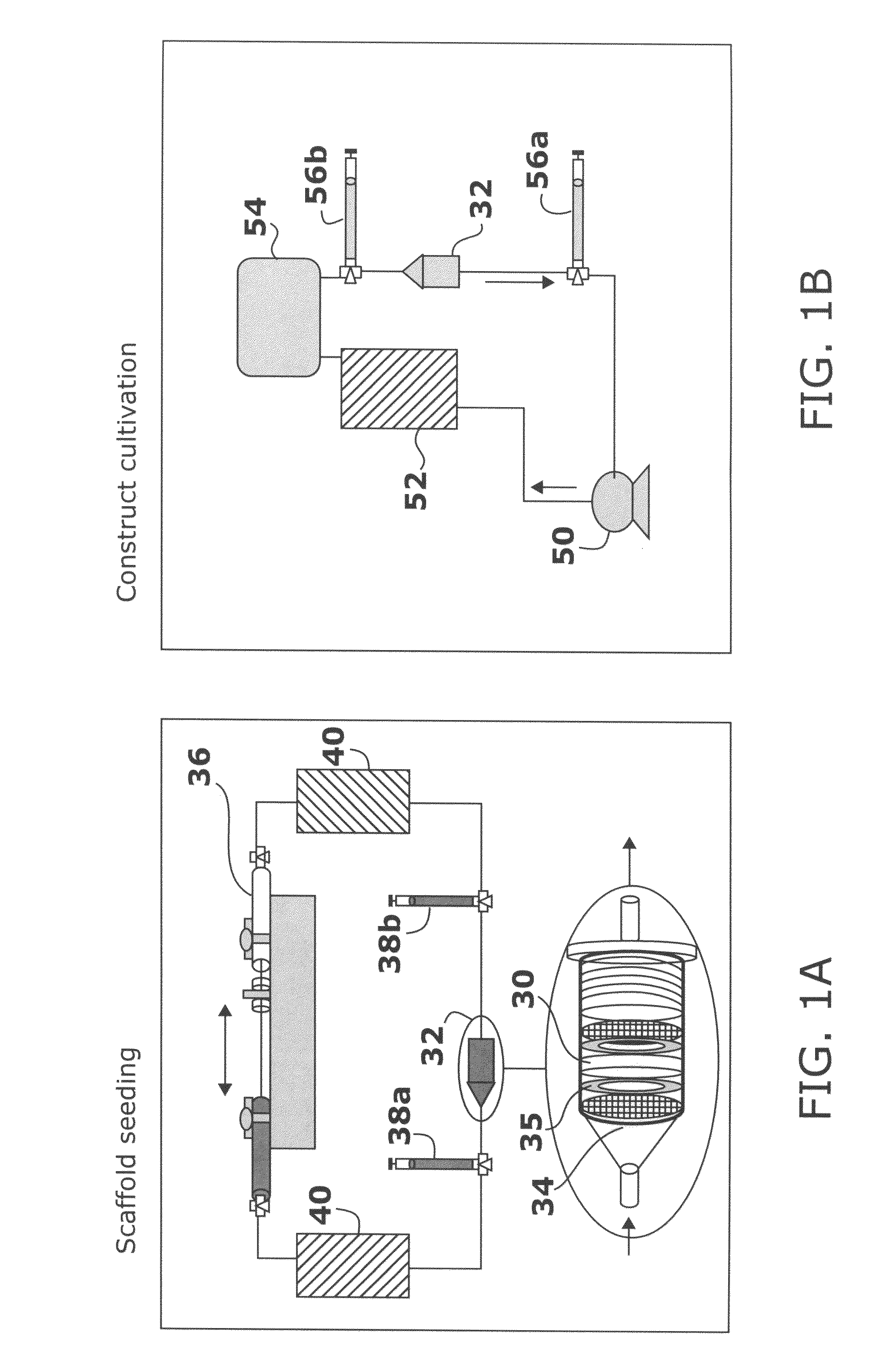
[0153]Cardiac Myocyte Preparation. Primary cultures of cardiac myocytes were prepared by enzymatic digestion of ventricles obtained from neonatal (2 day old) Sprague-Dawley rats (Taconic), as previously described (M. Toraason et al., Toxicology, 1989, 56: 107-113, the contents of which are incorporated herein by reference). Briefly, ventricles (n=50, 5 litters in 3 independent studies) were incubated with 0.1% trypsin overnight and dissociated in four to five sequential steps using 0.1% collagenase.

[0154]Cell-Seeded Construct Preparation. Isolated cardiac myocytes were suspended in Matrigel® (106 cells / 5 μL) and seeded onto Ultrafoam™ collagen sponge scaffold (6×8×1.5 mm) at the density of 0.8×10⁸ cells / cm3.

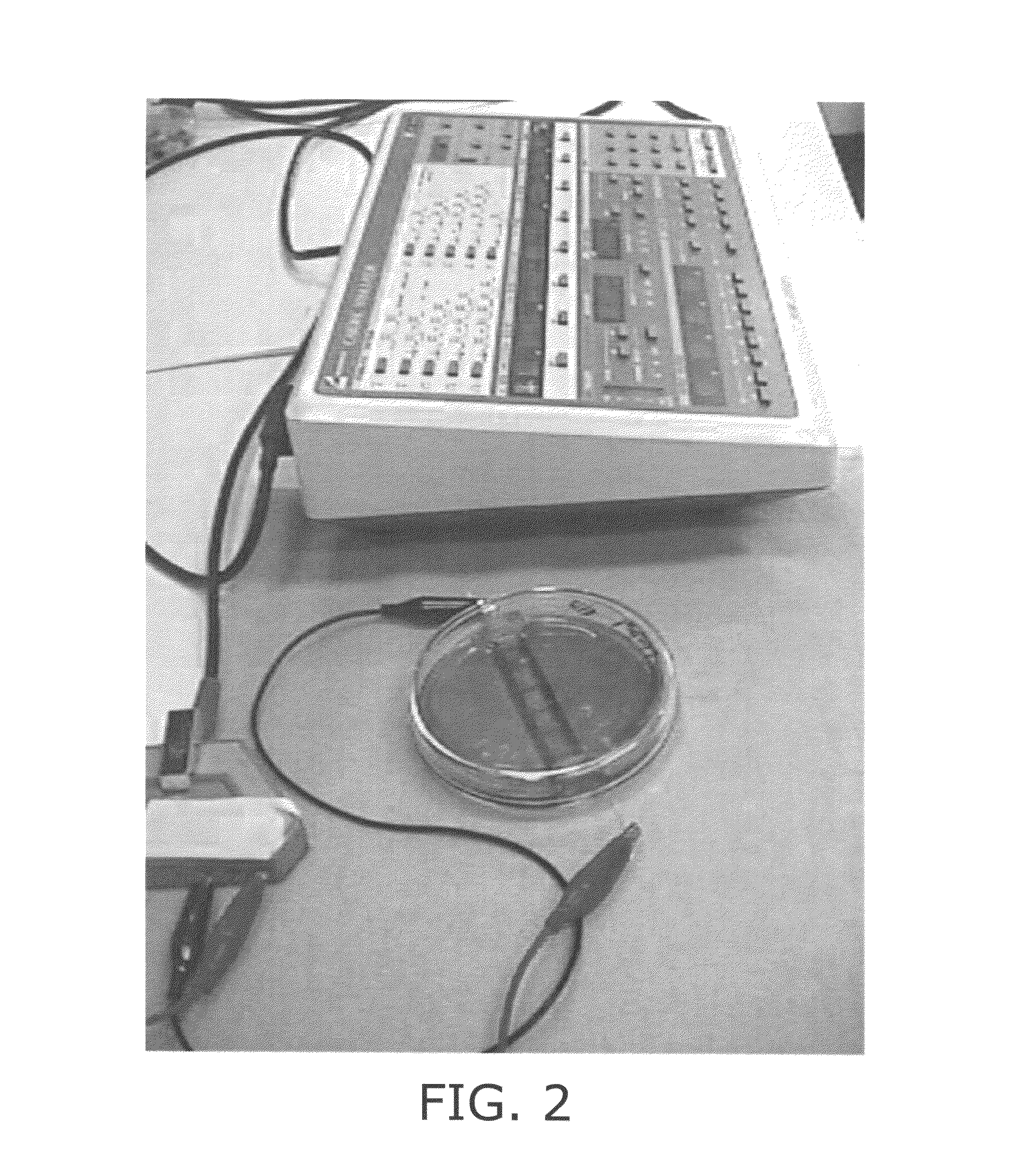
[0155]Cultivation of Cell-Seeded Construct. After 3 days of cultivation, the engineered tissue was transferred to a chamber consisting of a 100 mm glass Petri dish fitted with two ¼″ diameter carbon rods (stimulating electrodes) c...

example 2

Histological and Immunohistochemical Assessments

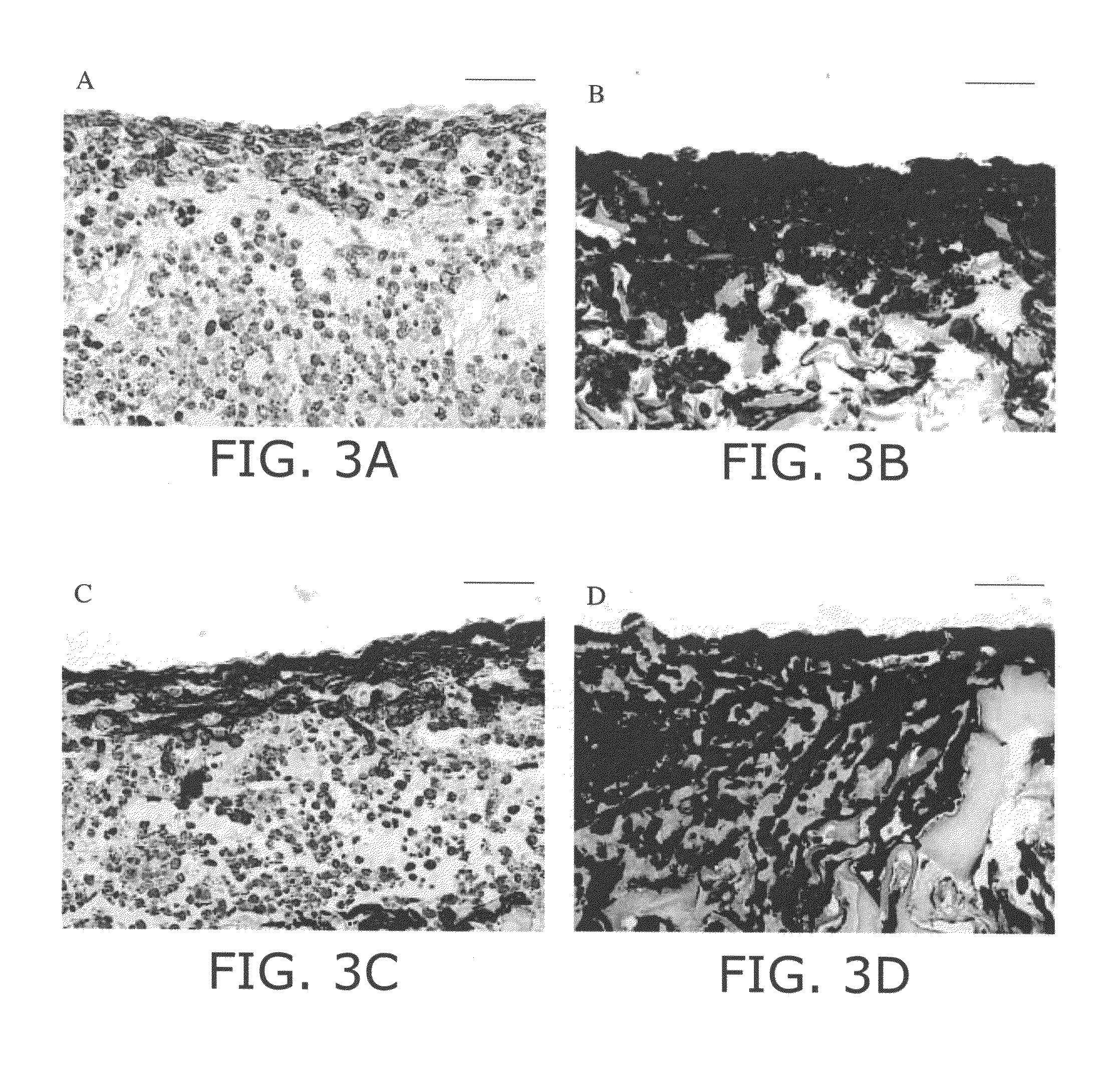
[0157]General Evaluation. Cardiac muscle constructs obtained as described in Example 1 in the presence or in the absence of electrical stimulation were rinsed in PBS, and immersed in 10% neutral buffered formalin (Sigma-Aldrich). Samples were embedded in paraffin, sectioned at 5 μm, and stained with hematoxylin and eosin (H+E) for general evaluation.

[0158]Tissue Architecture and Cell Distribution. Staining for cardiac troponin I, sarcomeric α-actin, α-myosin heavy chain (α-MHC), β-myosin heavy chain (β-MHC) (myocyte-specific contractile proteins) and connexin-43 (Cx-43, gap junctional protein) was used to assess the fraction and distribution of cardiac myocytes in the constructs.

[0159]For immunohistochemical staining, cardiac tissue sections were depariffinized and antigen was retrieved by heat treatment for 20 minutes at 95° C. in decloaking chamber (Biocare Medical). Subsequently, endogenous peroxidase activity was quenched by incuba...

example 3

Biochemical and Molecular Assays

[0163]DNA and Protein Assays. Samples for DNA, total protein, and Western blot analyses are homogenized in buffer (1 M NH4OH / 2% Triton X-100, 0.04 mL / mg wet wt of sample) for 1 minute. DNA is measured fluorimetrically by Hoescht binding, and total protein is measured by a commercially available kit (Bio-Rad) as previously described (N. Bursac et al., Am. J. Physiol. Heart Circ. Physiol. 1999, 277: H433-H444; R. L. Carrier et al., Biotechnol. Bioeng. 1999, 64: 580-589).

[0164]Protein Analysis. The protein expression levels of creatine kinase-MM (CK-MM) and myosin heavy chain (MHC), troponin I and connexin-43 in constructs are quantified as markers of cellular differentiation. Creatine kinase, a dimer of M- or B-type subunits, plays a vital role in the maintenance of cytosolic ATP (L. H. Opie, The Heart Physiologyfrom Cell to Circulation, L. Opie (Ed.), Lippincott-Raven: Philadelphia, 1998, pp. 295-342), whereas MHC is involved in the generation of contr...


PUM

PropertyMeasurementUnit
depthsaaaaaaaaaa
thicknessaaaaaaaaaa
thickaaaaaaaaaa

Abstract

The present invention provides new methods for the in vitro preparation of bioartificial tissue equivalents and their enhanced integration after implantation in vivo. These methods include submitting a tissue construct to a biomimetic electrical stimulation during cultivation in vitro to improve its structural and functional properties, and / or in vivo, after implantation of the construct, to enhance its integration with host tissue and increase cell survival and functionality. The inventive methods are particularly useful for the production of bioartificial equivalents and / or the repair and replacement of native tissues that contain electrically excitable cells and are subject to electrical stimulation in vivo, such as, for example, cardiac muscle tissue, striated skeletal muscle tissue, smooth muscle tissue, bone, vasculature, and nerve tissue.

Description

[0001]This application claims priority from U.S. Provisional Application No. 60 / 480,214, filed Jun. 20, 2003, the entire contents of which are incorporated herein by reference. To the extent that the incorporated contents conflict with this application, this application shall prevail.GOVERNMENT INTERESTS[0002]The work described herein was partly supported by the National Aeronautics and Space Administration (NASA, Grant No. NCC8-174). The United States government may have certain rights in the invention.BACKGROUND OF THE INVENTION[0003]Cardiovascular diseases are responsible for a preponderance of health problems in the majority of the developed countries as well as in many developing countries. Heart disease and stroke, the principal components of cardiovascular disease, are the first and third leading cause of mortality in the U.S., accounting for nearly 40% of all deaths (Heart and Stroke Statistical Update, American Heart Association 2002). Cardiovascular diseases also include c...

Claims


Application Information

Patent Timeline
Patent Type & Authority Patents(United States)
IPC IPC(8): C12N5/00C12N5/02C12N5/07A61F2/00A61NC12N5/077
CPCC12N5/0658C12N2502/02C12N2529/00C12N2502/1358
Inventor RADISIC, MILICAPARK, HYOUNGSHINLANGER, ROBERTFREED, LISAVUNJAK-NOVAKOVIC, GORDANA
Owner MASSACHUSETTS INST OF TECH
Why Eureka
Try Eureka