
Compositions comprising nanostructures for cell, tissue and artificial organ growth, and methods for making and using same

a technology of cell and tissue, and nanostructures, applied in the direction of micromachined delivery, isotope separation, capsule delivery, etc., can solve the problems of inability to investigate such titanium oxide nanotubes for bone growth or other bio applications, the size, spacing and height of nanotubes are not always easy to control, and the dimension regime of approximately 100 nm is too small to accommodate. , to achieve the effect of accelerating bone formation

Active Publication Date: 2013-04-09
RGT UNIV OF CALIFORNIA
View PDF19 Cites 63 Cited by
  • Summary
  • Abstract
  • Description
  • Claims
  • Application Information

AI Technical Summary

Benefits of technology

The patent describes a way to grow self-organized TiO2 nanotube arrays on titanium metal or alloy substrates to promote cell proliferation. The base material can be pure titanium or titanium-based alloys. The nanotube arrays can be used for various applications such as accelerating bone growth, promoting cell proliferation and harvesting, treating diseases with sustained release of pharmaceutical compositions, and diagnosing diseased or epidemic cells.

Problems solved by technology

While the fabrication of vertically aligned TiO2 nanotubes on Ti substrate was demonstrated by anodization process, an investigation of such titanium oxide nanotubes for bone growth or other bio application has not been attempted.
The structure of the anodized TiO2 nanotube array, such as the diameter, spacing and height of nanotubes, is not always easy to control during the electrochemical anodization process of pore formation.
While a portion of filopodia, the thin branches of growing cells, can get into such a small pores and enhance cell adhesion / growth, the approximately 100 nm regime of dimension is too small to accommodate the main part of typical osteoblast and many other cells as these have a much larger dimension of micrometers.
However, the fatal drawback of these currently available coating techniques is that such a flat and continuous coatings tend to fail by fracture or de-lamination at the interface between the implant and the coating as an adhesion failure, or at the interface between the coating and the bone, or at both boundary interfaces.
Although liver transplantation is an optional treatment method, there is a very limited supply of donor organs, and the medical and associated costs for the transplant procedure and post-operation immunosuppressive drug therapy are considerable.
However, there are a number of known drawbacks about long-term liver cell culture as some loss of liver function is frequently observed.
So far, there has been no successful means of proliferating healthy liver parenchymal cells.
Even though many of these experimental approaches have demonstrated an extended viability of hepatocyte and the stability of liver specific function under in vivo environment, they are still not satisfactory enough for practical applications.
However, there are still problems about the nature of non-liver derived cells.
The viability and functional activities of co-cultured hepatic primary cell can be prolonged in vitro, but primary cell proliferation is limited or absent in these system, which is a critical flaw.
Even though several reports indicate that non-parenchymal liver cells may express functions similar to hepatocytes, the nature of non-liver derived cells co-cultured with liver primary cells has not been established unequivocally.
One of the major roadblocks to the development of an effective artificial liver device is the lack of a satisfactory liver cell line that can provide the functions of a liver.

Method used


Image

  • Compositions comprising nanostructures for cell, tissue and artificial organ growth, and methods for making and using same
  • Compositions comprising nanostructures for cell, tissue and artificial organ growth, and methods for making and using same
  • Compositions comprising nanostructures for cell, tissue and artificial organ growth, and methods for making and using same

Examples


example 1

Bone- and Cell-growth Promoting Nanostructures

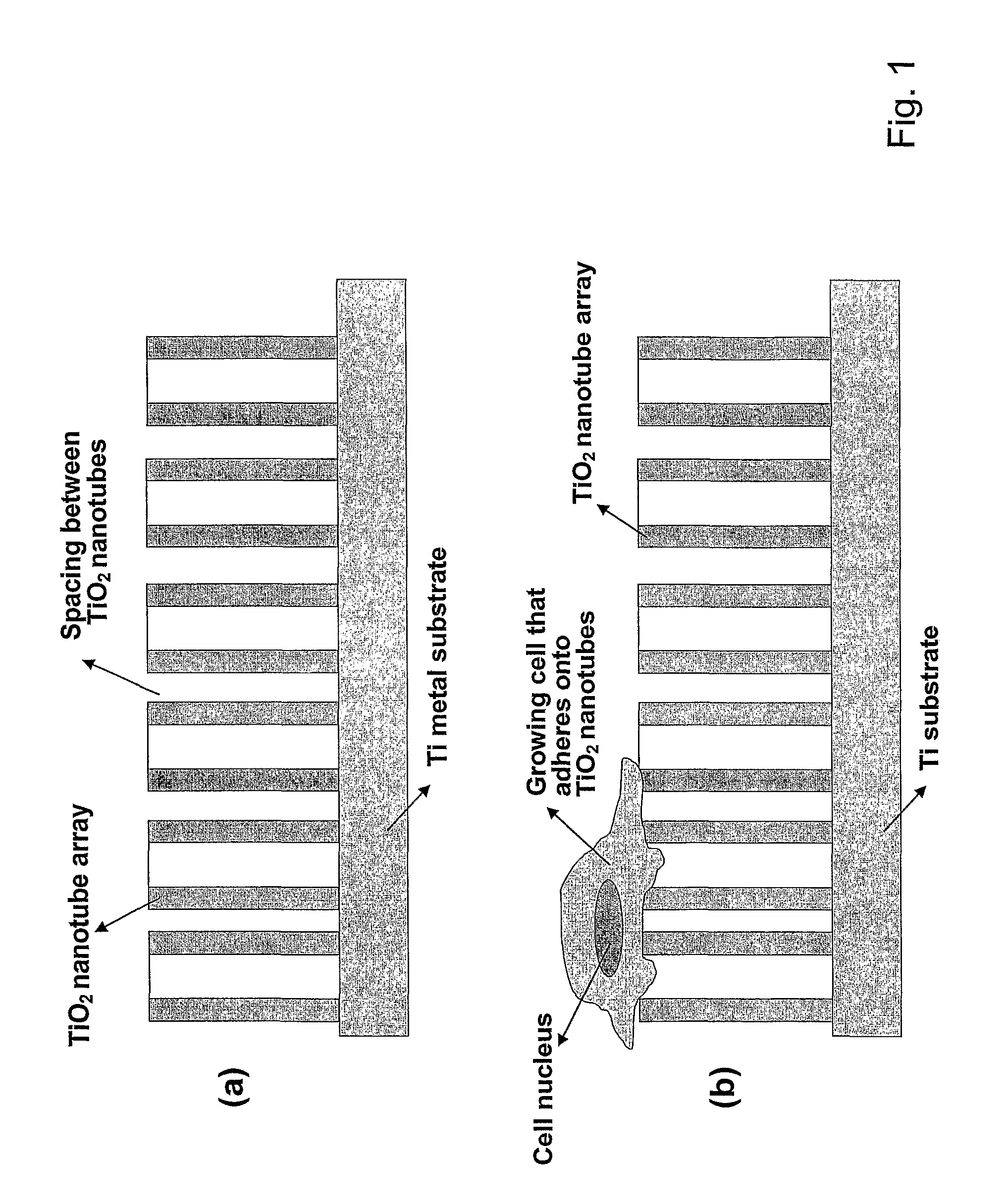
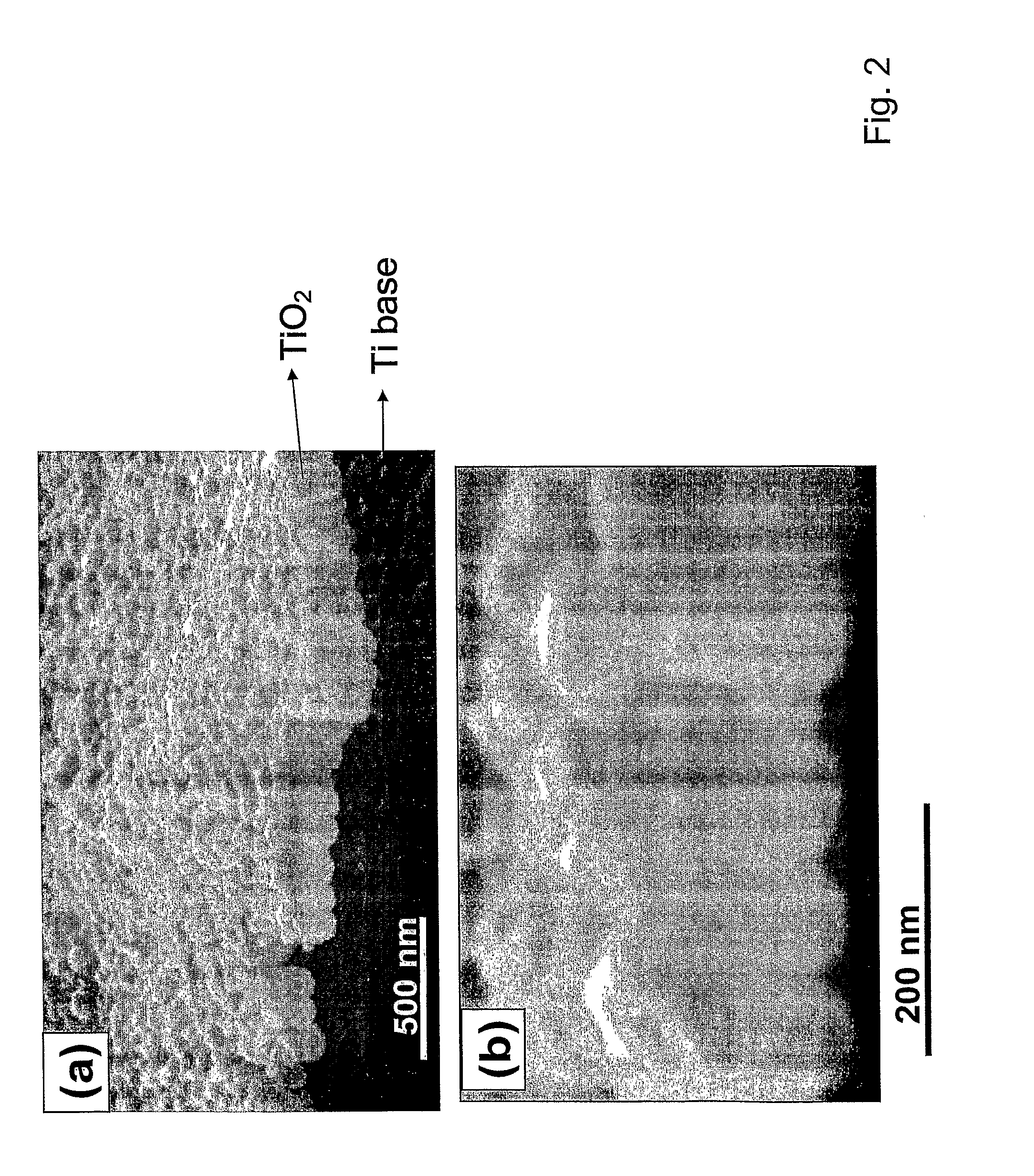
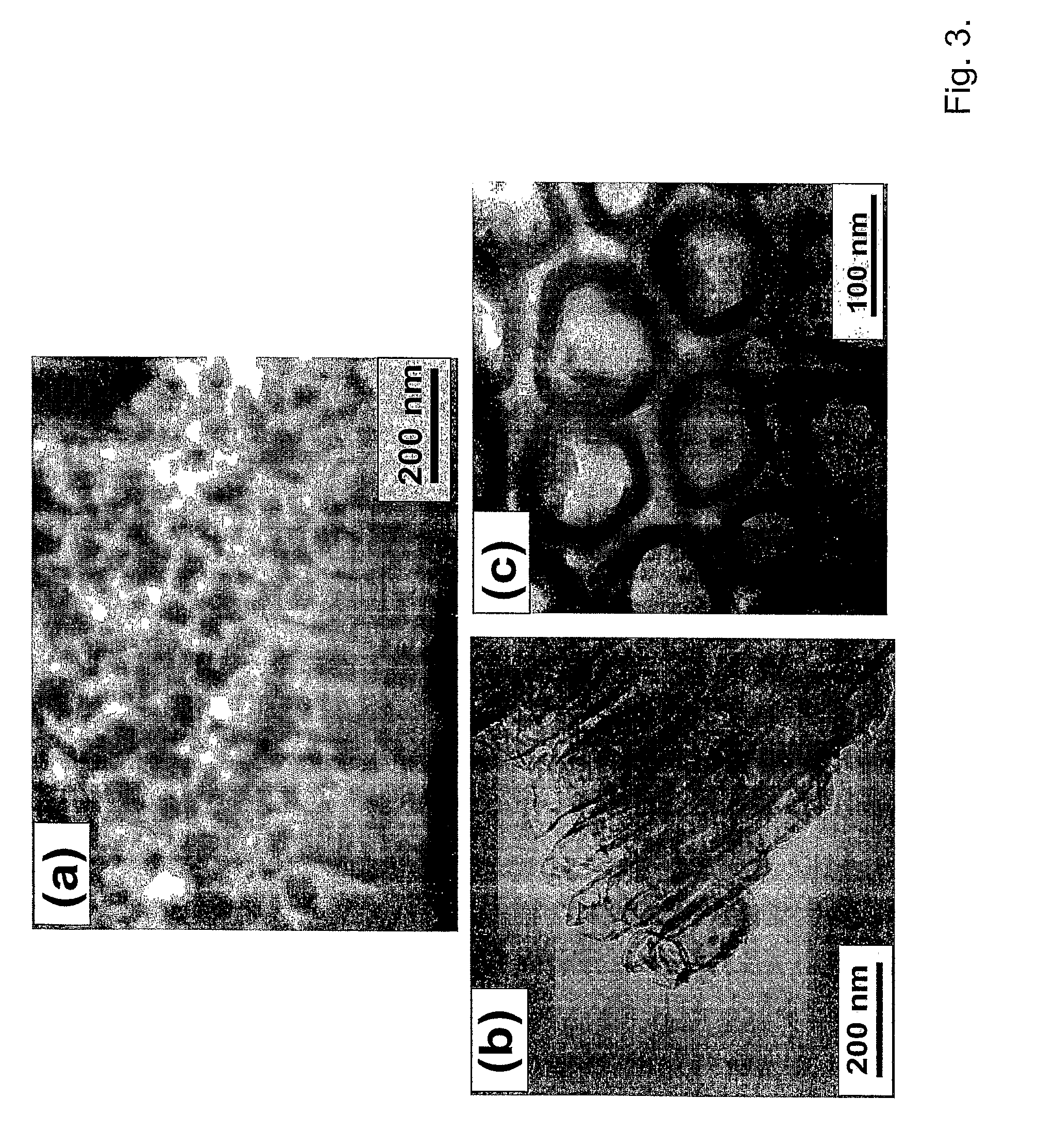
[0067]An example of an exemplary bone- and cell-growth promoting nanostructure of the invention is shown in FIGS. 2 and 3. These exemplary structures of the invention are vertically aligned, biocompatible TiO2 with a typical dimension of the hollow nanotubes as shown as being approximately (about) 100 nm outer diameter and approximately 70 nm inner diameter, with approximately 15 nm in wall thickness, and approximately 250 nm in height.

[0068]The exemplary TiO2 nanotube array structure shown in FIG. 1-3 was fabricated by an exemplary anodization technique using a Ti sheet (0.25 mm thick, 99.5% purity) which is electrochemically processed in a 0.5% HF solution at 20 V for 30 min at room temperature. A platinum electrode (thickness: 0.1 mm, purity: 99.99%) was used as the cathode. To crystallize the deposited amorphous-structured TiO2 nanotubes into the desired anatase phase, the specimens were heat-treated at 500° C. for 2 hrs. In one aspe...

example 2

Nanofiber-like or Nanoribbon-like Structures

[0071]On exposure of the TiO2 nanotubes to a 5 mole NaOH solution at approximately 60° C. for 60 minutes, it has been found that an additional, extremely fine, and predominantly nanofiber-like or nanoribbon-like structure of sodium titanate compound is introduced on the very top of the TiO2 nanotubes as shown in FIG. 5(a). In this example, preferential occurrence of nanofibers at the top of nanotubes is presumably because of the nanotube contact with NaOH solution above and also possibly due to the surface-tension-related difficulty of NaOH solution getting into nanopores within and in-between TiO2 nanotubes, as illustrated in FIG. 5(b). Compositional analysis by EDXA (energy dispersive x-ray analysis) in SEM confirms the presence of Na, Ti and O after the exposure of TiO2 nanotubes to NaOH. The sodium titanate so introduced exhibits an extremely fine-scale nanofiber configuration with a dimension of approximately 8 nm in average diameter ...

example 3

Osteoblast Cell Growth on Nanotubes of the Invention

[0076]In order to estimate the effect of having an extremely fine nanostructure such as the vertically aligned TiO2 nanotubes on cell growth behavior, an osteoblast cell growth on TiO2 nanotubes was performed. The results demonstrate (indicate) that the introduction of nanostructure significantly improves bioactivity of implant and enhances osteoblast adhesion and growth. An adhesion of anchorage-dependent cells such as osteoblasts is a necessary prerequisite to subsequent cell functions such as synthesis of extracellular matrix proteins, and formation of mineral deposits. In general, many types of cells beside the osteoblast cells remain healthy and grow fast if they are well-adhered onto a substrate surface, particularly a nanostructure surface of this invention, while the cells not adhering to the surface tend to stop growing.

[0077]All the experimental specimens (0.5×0.5 cm2) used for cell adhesion assays were sterilized by auto...


PUM

PropertyMeasurementUnit
outer diameteraaaaaaaaaa
heightaaaaaaaaaa
vertical alignment angleaaaaaaaaaa

Abstract

The invention provides articles of manufacture comprising biocompatible nanostructures comprising nanotubes and nanopores for, e.g., organ, tissue and / or cell growth, e.g., for bone, kidney or liver growth, and uses thereof, e.g., for in vitro testing, in vivo implants, including their use in making and using artificial organs, and related therapeutics. The invention provides lock-in nanostructures comprising a plurality of nanopores or nanotubes, wherein the nanopore or nanotube entrance has a smaller diameter or size than the rest (the interior) of the nanopore or nanotube. The invention also provides dual structured biomaterial comprising micro- or macro-pores and nanopores. The invention provides biomaterials having a surface comprising a plurality of enlarged diameter nanopores and / or nanotubes.

Description

FIELD OF THE INVENTION[0001]The present invention provides articles of manufacture comprising biocompatible nanostructures comprising nanotubes and nanopores for, e.g., organ, tissue and / or cell growth, e.g., for bone, tooth, kidney or liver growth, and uses thereof, e.g., for in vitro testing of drugs, chemicals or toxins, or as in vivo implants, including their use in making and using artificial tissues and organs, and related diagnostic, screening, research and development and therapeutic uses.BACKGROUND OF THE INVENTION[0002]It is known that the nano-scaled materials exhibit extraordinary electrical, optical, magnetic, chemical and biological properties, which cannot be achieved by micro-scaled or bulk counterparts. The development of nano-scaled materials has been intensively pursued in order to utilize such properties for various technical applications including biomedical and nano-bio applications.[0003]Ti and Ti alloys are corrosion resistant, light, yet sufficiently strong ...

Claims


Application Information

Patent Timeline
Patent Type & Authority Patents(United States)
IPC IPC(8): A61F2/00
CPCA61K9/0009A61K9/0024A61K9/0097A61L27/06A61L27/306A61L27/54A61L27/56C12M23/34A61L2300/254A61L2300/256A61L2300/406A61L2300/414A61L2300/416A61L2300/43A61L2300/624A61L2400/12C12M25/00C12M23/20A61M37/0092A61M2037/0007
Inventor JIN, SUNGHOOH, SEUNGHAN
Owner RGT UNIV OF CALIFORNIA
Learn More