Organ-specific proteins and methods of their use

a technology of organ-specific proteins and methods, applied in the field of organ-specific proteins and polynucleotides, can solve the problems of reducing patient survival, preventing early detection and diagnosis, and current diagnostic assays that may not detect disease,

Active Publication Date: 2013-11-19
INSTITUTE FOR SYSTEMS BIOLOGY +1
View PDF8 Cites 142 Cited by
  • Summary
  • Abstract
  • Description
  • Claims
  • Application Information

AI Technical Summary

Problems solved by technology

Current diagnostic assays may not detect disease until it has progressed to a stage where it is too late for effective treatment.
If cancer is diagnosed at later or advanced stages, effective treatment may be difficult or impossible and lead to reduced patient survival.
In general, current diagnostic assays have severe limitations that prevent early detection and diagnosis.
In the context of blood protein diagnostics the major impediment to use in the early detection of disease is that most proteins are not disease-specific in that multiple organs synthesize them and different diseases may perturb their expression in different ways.
Moreover, the specific proteins that are released in the disease state that are markers of the disease may be difficult to identify or to measure because of the enormous dynamic range of protein expression in the blood and because of the enormous protein complexity in the blood.
Most of these abundant blood proteins are not useful diagnostic markers.
Additionally, many proteins that are low abundance proteins are not indicative of disease.
Distinguishing between the low abundance proteins that indicate disease from the low abundance proteins that are found in the normal cellular state is a major challenge to modern protein diagnostics.
A major obstacle in diagnostic protein analysis of the blood is the numerous blood proteins and an inability by current methods to distinguish proteins from one another.
Determining which blood proteins are predictive of disease at the earliest stages is very difficult at best, because the diagnostician must distinguish which low abundance protein is a marker of disease within the mass of proteins that are circulating in the blood.
Different approaches for identifying blood proteins are known in the art and have been used with varying and limited degrees of success.
In particular, two-dimensional gel electrophoresis (2-DE) has been used for analysis of proteomic patterns in blood, but it is difficult to resolve large numbers of proteins such as are expressed in the average cell (up to 10,000 proteins).
Moreover, 2-DE is incapable of identifying low abundance proteins without enrichment techniques.
Another method known in the art for blood protein diagnostics is capillary isoelectric focusing electrophoresis (CGE) although, the lack of reproducibility of protein patterns limits its use (Corthals, G. L., et al.
Consequently, protein pattern analysis using techniques such as 2-DE, CGE and other similar techniques cannot generally be used for the analysis of blood proteins due to the inability to detect very low abundance proteins, irreproducible gel patterns, and the inability to quantify or identify individual spots (e.g., proteins).
Further, the ability to extend these techniques to reproducible, consistent, easy to use and accurate high throughput diagnostic assays has been extremely limited.
Thus, current assays that detect proteins do not provide the accuracy to use levels of blood (or other biological fluid or tissue) proteins, polypeptides or nucleic acids to monitor health and disease.

Method used


Image

  • Organ-specific proteins and methods of their use
  • Organ-specific proteins and methods of their use
  • Organ-specific proteins and methods of their use

Examples


example 1

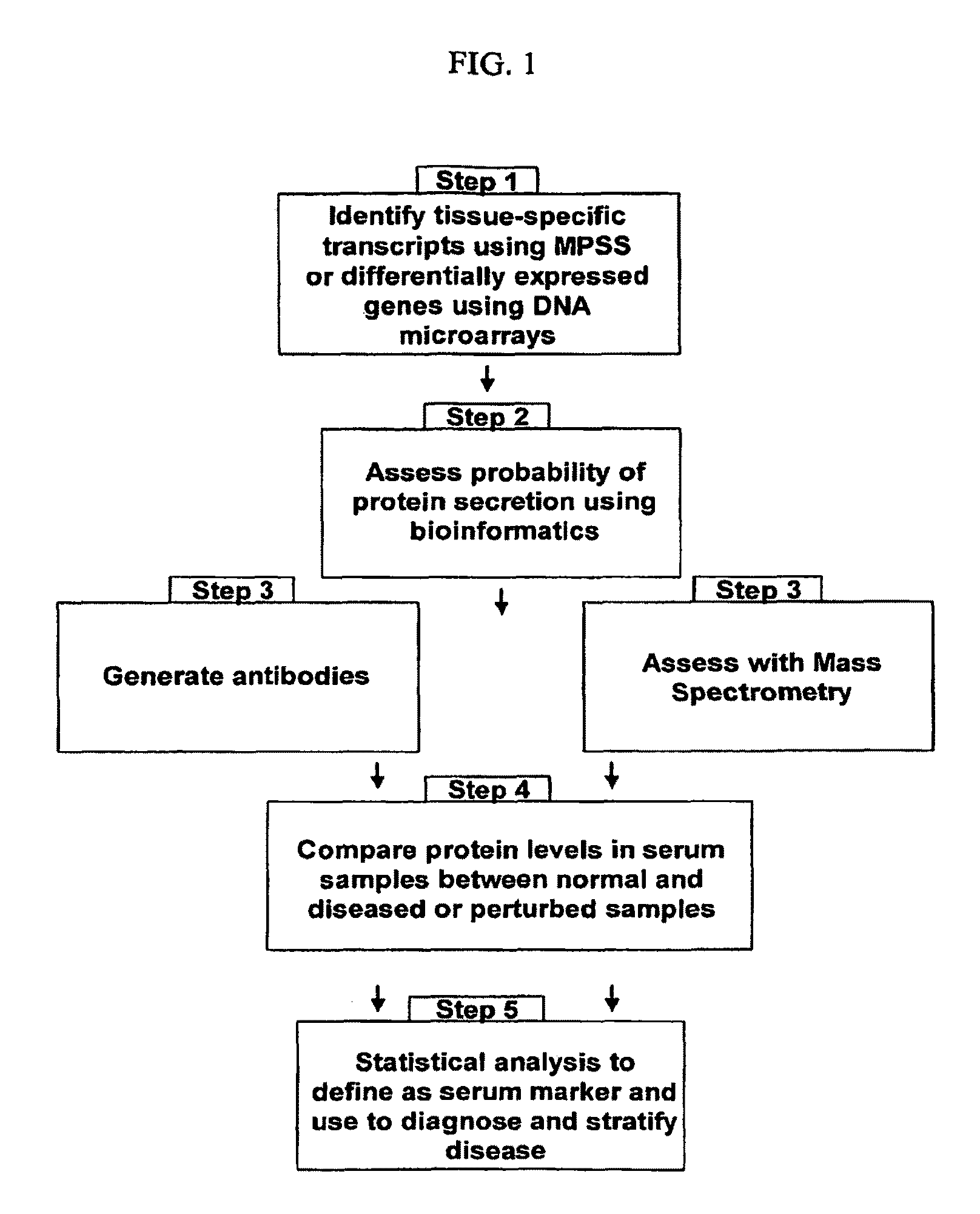
Identification of Organ-Specific Proteins by Analysis of MPSS Data

[0696]This example shows the identification of organ-specific protein sets from 32 normal, healthy organs.

1. Normalized MPSS Dataset

[0697]The normalized MPSS data used in this study was previously described (Jongeneel, et al., Genome (2005), 15:1007-1014) but re-annotated by Solexa (Hayward, Calif., USA) to the new UniGene database. The data contained a total of 391,669 MPSS sequence tags, their gene annotation, their expression levels in 32 different tissues as specified by their transcription per million (tpm) and the associated standard deviation (SD), and information on the sequence tags such as significance, selected step, class, and global replication. This dataset was used to identify organ-specific proteins as described below.

Identification of Organ-Specific MPSS Tags

[0698]Assume the expression (in tpm) and the associated SD of a MPSS sequence tag in the 32 tissues were {(Xi,σi)}, where i=1, 2, . . . , 32 repr...

example 2

Identification of Organ-Specific Proteins in Human Serum Using Mass Spectrometry

[0723]This experiment demonstrates the process of identifying organ-specific proteins in a normal sample of blood serum from a healthy, human volunteer. For normal control serum, venous blood samples were drawn from a fasted, human volunteer. Samples were collected with minimal stasis in evacuated serum separator tubes. After at least 30 min, but within 2 hours, the tubes were centrifuged at 23° C. for 15 minutes at 1,200 g and serum was stored in plastic vials at −80° C. To reduce sample complexity, plasma was passed over a column containing antibodies to the most abundant proteins. In this example, an affinity column was used to remove albumin, IgG, IgA, anti-trypsin, transferrin, and haptoglobin; however, affinity columns with an expanded repertoire could also be used. Since most proteins found on the cell surface or secreted from cells are glycoproteins, and can be isolated via the glycopeptide captu...

example 3

Verification and Quantification of Serum Proteins Using Enzyme-Linked Immunosorbant Assay (ELISA)

[0730]Blood serum tests to detect and monitor proteins were developed using an enzyme-linked immunosorbent assay (ELISA). The assay system utilized two antibodies directed against different antigenic regions of the candidate protein. A monoclonal antibody directed against a distinct antigenic determinant on the intact candidate protein was used for solid phase immobilization on the microtiter wells. A detection antibody conjugated to horseradish peroxidase (HRP) or fluorescence tag recognized the candidate protein within different region of the same protein. The candidate protein reacted simultaneously with the two antibodies, resulting in the protein being sandwiched between the solid phase and detection antibody. The detection antibody was visualized by colorimetric fluorescence analysis.

[0731]In the case of peptides detected in blood, the specific peptide was further enriched from pep...


PUM


Abstract

The present invention relates generally to methods for identifying and using organ-specific proteins and transcripts. The present invention further provides compositions comprising organ-specific proteins and transcripts encoding the same, detection reagents for detecting such proteins and transcripts, and diagnostic panels, kits and arrays for measuring organ-specific proteins / transcripts in blood, biological tissue or other biological fluid.

Description

STATEMENT OF GOVERNMENT INTEREST[0001]This invention was made with government support under Grant Nos. P50 CA097186 and P01 CA085859 awarded by the National Cancer Institute. The government may have certain rights in this invention.SUBMISSION OF SEQUENCE LISTING ON COMPACT DISC[0002]The content of the following submission on compact discs is incorporated herein by reference in its entirety: A computer readable form (CRF) of the Sequence Listing on compact disc (file name: 655652003300Seqlist.txt, date recorded: Nov. 3, 2011, size: 135,150,157 bytes); a duplicate compact disc copy of the Sequence Listing (COPY 1) (file name: 655652003300Seqlist.txt, date recorded: Nov. 3, 2011, size: 135,150,157 bytes); and a duplicate compact disc copy of the Sequence Listing (COPY 2) (file name: 655652003300Seqlist.txt, date recorded: Nov. 3, 2011, size: 135,150,157 bytes).STATEMENT REGARDING TABLES SUBMITTED ON CD-ROM[0003]Tables 1A-32A, 1B-32B, 36A-45A, 36B-45B, 47A-79A and 47B-79B associated wit...

Claims


Application Information

Patent Timeline
Patent Type & Authority Patents(United States)
IPC IPC(8): A61K49/00
CPCG01N33/6845G01N2800/00G01N2800/52A61P35/00G01N33/68
Inventor HOOD, LEROYBECKMANN, PATRICIA M.JOHNSON, RICHARDMARELLI, MARCELLOLI, XIAOJUN
Owner INSTITUTE FOR SYSTEMS BIOLOGY
Why Eureka
Try Eureka