Glucomannan scaffolding for three-dimensional tissue culture and engineering

a scaffolding and three-dimensional technology, applied in 3d culture, non-embryonic pluripotent stem cells, skeletal/connective tissue cells, etc., can solve the problems of limiting clinical application, inconformity of mechanical properties and behavior of seeded cells, and the inability to conduct glucomannan as a porous scaffold for tissue engineering applications, etc., to achieve the effect of promoting cell adhesion and proliferation

Active Publication Date: 2016-06-07
RGT UNIV OF CALIFORNIA
View PDF7 Cites 8 Cited by
  • Summary
  • Abstract
  • Description
  • Claims
  • Application Information

AI Technical Summary

Benefits of technology

[0009]It has been found that when a basic glucomannan scaffold is neutralized, the resulting neutralized glucomannan scaffold is useful as a matrix for three-dimensional (3D) cell culture and tissue engineering. The neutralized glucomannan scaffold has a pH that is suitable for efficient cell growth, and a porous structure that can permit diffusion of oxygen, nutrients, expressed products and cellular waste. Moreover, the neutralized glucomannan scaffold of the invention is amenable to surface modification to promote cell adhesion and proliferation. This naturally-derived biomaterial scaffold is thermally stable, non-toxic and biodegradable and may mimic the natural three-dimensional microenvironment of a wide range of cell types, such as osteoblasts, heptocytes, lymphocytes and stem cells.

Problems solved by technology

However, challenges with inconsistent mechanical properties and behavior of seeded cells limit their clinical application.
However, an exploration of developing glucomannan as a porous scaffold for tissue engineering applications has yet to be conducted.

Method used


Image

  • Glucomannan scaffolding for three-dimensional tissue culture and engineering
  • Glucomannan scaffolding for three-dimensional tissue culture and engineering
  • Glucomannan scaffolding for three-dimensional tissue culture and engineering

Examples


example 1

Preparation of Glucomannan Scaffold

[0082]Glucomannan powder (1-5 g) was dissolved in 100 ml of distilled water and stirred slowly for 5 min, then the solution was incubated at room temperature for 60 min. Calcium hydroxide solution (1.5%, 10 ml; Sigma-Aldrich, St. Louis, Mo., USA) was added to the glucomannan solutions and mixed vigorously for 1 min. Poly-L-lysine (Sigma-Aldrich) (0.001% to 1% w / v aqueous solution) was added to the mixture and heated to 125° C. in a decloaking chamber for 30 min. After cooling to room temperature, glucomannan gels were soaked in distilled water overnight. Glucomannan gels were placed in culture dishes and then frozen in a blast freezer for 30 min at less than or equal to −50° C. Water was sublimated using a Vitris model 50-SRC-5 Sublimator. The shelf temperature was 12° C. with a condenser temperature of less than or equal to −58° C., and the vacuum was maintained at 80-100 millitorr. The resulting glucomannan products were then packed in polyethyle...

example 2

Cell Growth on Neutralized Glucomannan Scaffold

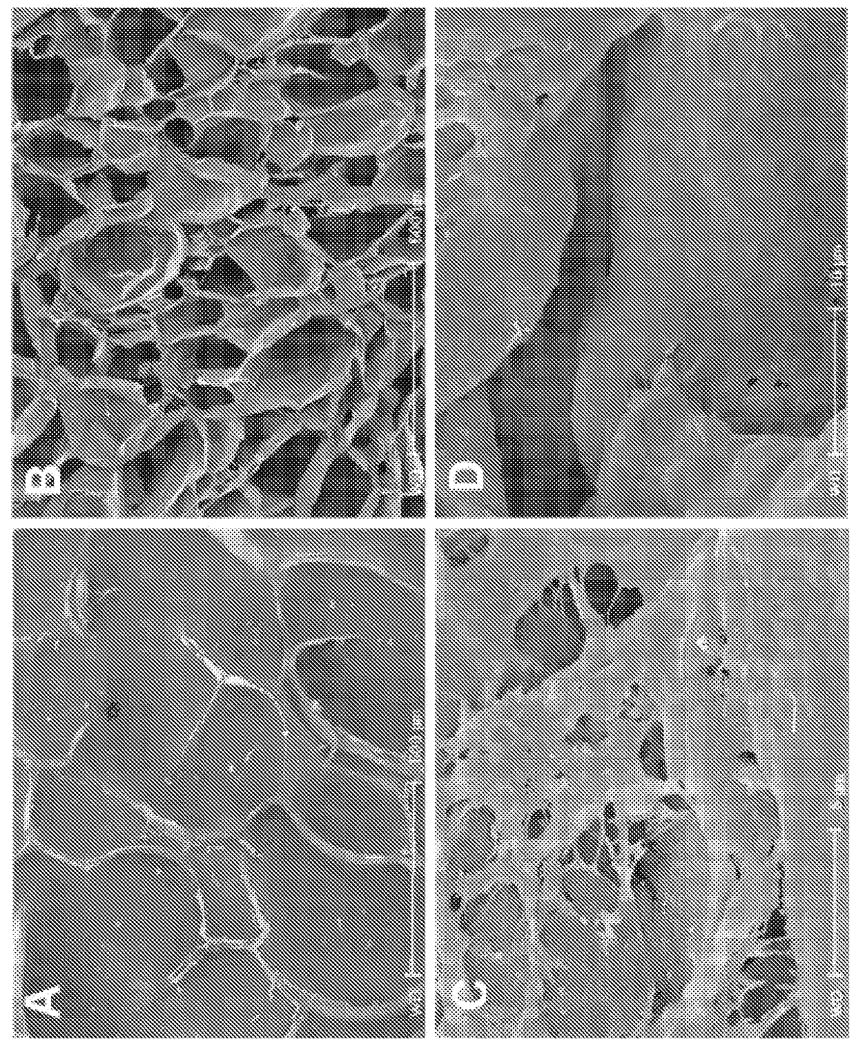
[0088]Preparation of Cells. For human mesenchymal stem cells (hMSC), bone marrow mononuclear cells (N=3, 1 female and 2 males, 20-40 yrs of age; Cambrex, East Rutherford, N.J., USA) were plated in 100 mm plates in α-MEM containing 20% fetal bovine serum (FBS), 1% L-glutamine, and 1% penicillin-streptomycin (Invitrogen, Carlsbad, Calif., USA) and incubated at 37° C. in 5% CO2. Plates were washed with phosphate-buffered saline (PBS) three times every other day until cells reached approximately 80% confluence. Cells were incubated with 0.25% trypsin-EDTA (Invitrogen) for 5 min at 37° C. Culture medium was added to the cells in at a 1:1 ratio of trypsin-EDTA in order to inactivate. Cells were replated in culture medium at 5×10³ cells / cm2. Cells were cultured up to 3 passages, cryopreserved using a controlled rate protocol at every passage, and stored in liquid nitrogen until use. For CD34+ cells, a subset of bone marrow mononuclear cells me...

example 3

Enzyme Digestion

[0092]Lyophilized endo-1,4 β-mannanase (Megazyme) or cellulase obtained from Aspergillus niger (Sigma-Aldrich) were dissolved in culture medium. Scaffolds seeded with hMSC were incubated in 0.5 units / ml, 5.0 units / ml, or 50 units / ml of endo-1,4 β-mannanase or cellulase overnight. Degradation of scaffolds was checked visually. Cells were counted once scaffolds were completely dissolved.


PUM

PropertyMeasurementUnit
pressureaaaaaaaaaa
temperatureaaaaaaaaaa

Abstract

The present invention provides a neutralized glucomannan scaffold capable of promoting cell growth and suitable for three-dimensional tissue culture and engineering. The present invention also provides methods for making and degrading the neutralized glucomannan scaffold. The present invention further provides a method of growing cells on a neutralized glucomannan scaffold.

Description

CROSS-REFERENCE TO RELATED APPLICATIONS[0001]This application is the U.S. National Stage Entry under §371 of International Application No. PCT / US2012 / 066982, filed Nov. 29, 2012, which claims priority to U.S. Provisional Application No. 61 / 564,553, filed Nov. 29, 2011, which is incorporated in its entirety herein for all purposes.BACKGROUND OF THE INVENTION[0002]Engineering biomaterials to repair damaged or diseased tissues such as cardiac, bone, liver, corneal and skin is an active branch of research in regenerative medicine. One approach being investigated is using cells combined with biomaterial constructs, or scaffolds, that facilitate cell growth and differentiation to create functional tissues in vitro that can be implanted. Three-dimensional (3D) tissue culture systems, which emulate key physical and molecular features of the extracellular microenvironment, provide tremendous advantages to tissue engineering.[0003]Biomaterials may be naturally-derived, such as protein- and po...

Claims


Application Information

Patent Timeline
Patent Type & Authority Patents(United States)
IPC IPC(8): C12N5/074C12N5/00C12N5/0775C08B37/00C08L5/00C08J9/28C08J9/36
CPCC12N5/0607C08B37/0087C08J9/28C08J9/36C08L5/00C12N5/0068C12N5/0663C08L89/06C08L89/00C08L77/04C08J2201/0484C08J2207/10C08J2305/00C12N2513/00C12N2533/70
Inventor LEE, C. CHANG I.
Owner RGT UNIV OF CALIFORNIA
Why Eureka
Try Eureka