Biological adhesive liposome preparation for eyes and preparation method thereof

A liposome preparation and bioadhesion technology, which is applied in the directions of liposome delivery, medical preparations of inactive ingredients, and drug combinations, etc., can solve the problem of poor compliance of semi-solid dosage forms, low ocular bioavailability, and difficulty in Patient acceptance and other issues to achieve the effect of improving ocular bioavailability, improving bioavailability, and promoting ocular absorption

Inactive Publication Date: 2010-03-17
FUDAN UNIV
View PDF0 Cites 6 Cited by
  • Summary
  • Abstract
  • Description
  • Claims
  • Application Information

AI Technical Summary

Problems solved by technology

[0006] The purpose of the present invention is to overcome the short residence time of common eye drops in the prior art in the conjunctival sac, resulting in low ocular bioa

Method used


Image

  • Biological adhesive liposome preparation for eyes and preparation method thereof
  • Biological adhesive liposome preparation for eyes and preparation method thereof
  • Biological adhesive liposome preparation for eyes and preparation method thereof

Examples


Embodiment 1

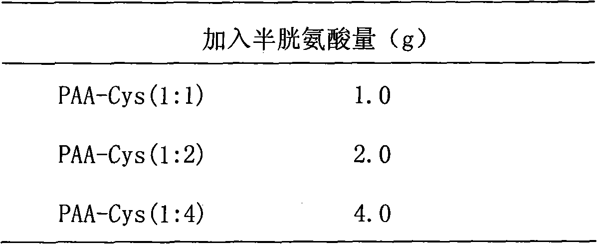
[0026] Preparation, separation and purification of cysteine-polyacrylic acid (Cys-PAA) complex: Take three parts of 1g PAA and add it to 100ml water, adjust the pH value to 7.2-7.5 with 2M NaOH, and disperse it in water. 3.8 g of 1-ethyl-3-(3-dimethylaminopropyl)carbodiimide hydrochloride (EDAC) was added each to make the concentration 200 mM, and stirred at room temperature for 45 minutes. Use 5M HCl to adjust the pH value to 4-5, fill with nitrogen for 15 minutes, then add Cys, fill with nitrogen and stir at room temperature for 3 hours. The amount of cysteine ​​added to each of the three parts is shown in Table 1.

[0027] Table 1

[0028]

[0029] The product was isolated by dialysis. The reaction solution was transferred to a dialysis bag, and dialyzed in 2000 ml of dialysis fluid at 4° C. in the dark. The first dialysate was 1 mM HCl containing 2 μM EDTA. The second dialysate was 1 mM HCl, containing 2 μM EDTA, and containing 1% NaCl, and the same medium was dialy...

Embodiment 2

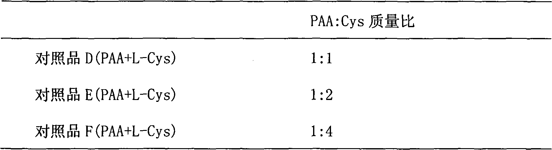
[0035]Determination of sulfhydryl content in Cys-PAA complex: The amount of sulfhydryl can be determined by DTNB method. Take 2mg each of the product and the reference substance and disperse them in 10ml of 0.5M phosphate buffer (PH8.0), respectively, take 0.5ml each, add 0.5ml of 0.03% 5,5-dimercapto-2,2-dinitrobenzoic acid (DTNB) solution, put it at room temperature for 2 hours, take 0.3ml and transfer it into a 96-well plate, put it into a microplate reader to measure the absorbance (absorbance value is set at 405nm). The cysteine ​​solution standard curve Y=163.38X+28.456 was obtained, and the sulfhydryl content in Cys-PAA was calculated based on this. The thiol content obtained after deducting the absorbance of the blank reference substance (D, E, F) is shown in Table 3:

[0036] table 3

[0037]

Embodiment 3

[0039] Evaluation of bioadhesion of PAA-Cys complex: Adult rats were taken, sacrificed after anesthesia, dissected, and the small intestinal mucosa was collected and stored at -20°C until use.

[0040] Take appropriate amounts of PAA-Cys (1:1), PAA-Cys (1:2), PAA-Cys (1:4), and PAA respectively to make a solution containing 0.2% PAA. Then it was divided into two parts, one was adjusted to neutral pH with 2M NaOH, and the other was adjusted to pH 3 with 5M HCl. Also adjust the pH value of pure water to neutral and 3. Take 0.2ml of each of the prepared above-mentioned solutions and drop them on the surface of the small intestinal mucosa, measure the adhesion force (mg) at 10 minutes and 30 minutes with a torsion balance, and repeat the measurement 6 times for each sample. The obtained adhesion results are shown in Table 4:

[0041] Table 4 (Neutral)

[0042]

[0043] (pH3)

[0044]

[0045] (Net Adhesion (mg))

[0046]

[0047]

[0048] The results showed that t...


PUM


Abstract

The invention belongs to the field of medicinal preparations, and relates to liposome for eyes and a preparation method thereof. In order to overcome the defects that common eye drops have short residence time in the conjunctival sac to cause low bioavailability at the eyes and the semi-solid dosage form has poor compliance and is not easy to be accepted by patients and the like in the prior art,the invention provides a biological adhesive liposome preparation for eyes. The biological adhesive liposome preparation for the eyes consists of the liposome, a liposome membrane modification material and a medicament wrapped in the liposome, wherein the surface of the liposome is modified with free mercapto, and a covalent binding disulfide bond can be formed by the free mercapto and a mucoprotein subdomain rich in cysteine on the surface of the eye to anchor the liposome on the surface of a mucous membrane and serve as a medicament store to slowly release the medicament in the conjunctivalsac and provide permanent driving force for the absorption of the medicament. The preparation is helpful for promoting the absorption of the medicament at the eyes, and can improve the bioavailabilityof the medicament.

Description

technical field [0001] The invention belongs to the field of pharmaceutical preparations, and relates to an ophthalmic liposome preparation and a preparation method thereof. The surface of the liposome is modified with free sulfhydryl groups, which can form a covalently bonded disulfide bond with the cysteine-rich subdomain of the ocular surface mucin, anchor the liposome on the mucosal surface, and act as a drug storage agent. The depot releases the drug slowly in the conjunctival sac, providing a sustained driving force for drug absorption. Background technique [0002] The eye is a highly sensitive organ with multiple protective mechanisms, so the design of an ocular drug delivery system is extremely challenging. Due to the rapid elimination of the medicinal solution dripped into the conjunctival sac and the bio-barrier effect of the cornea, less than 10% of the medicine in conventional eye drops can pass through the cornea and reach the eye to exert a therapeutic effect...

Claims


Application Information

Patent Timeline
IPC IPC(8): A61K9/127A61K47/32A61P27/02
Inventor 魏刚陆伟跃李翀谢操
Owner FUDAN UNIV
Why Eureka
Try Eureka