Preparation method of drug-loaded nanofiber medical dressing

A nanofiber and drug technology, applied in medical science, bandages, absorbent pads, etc., can solve the problems of iatrogenic pollution, damage to new tissue, wound adhesion, etc., and achieve accelerated healing, good absorption, and conducive to growth and regeneration Effect

Inactive Publication Date: 2011-12-07
HANGZHOU INST OF ADVANCED MATERIAL BEIJING UNIV OF CHEM TECH
View PDF2 Cites 28 Cited by
  • Summary
  • Abstract
  • Description
  • Claims
  • Application Information

AI Technical Summary

Problems solved by technology

[0005] At present, there are many inconveniences in the use of medical dressings. Some dressings use non-degradable polyacrylonitril

Method used


Image

  • Preparation method of drug-loaded nanofiber medical dressing
  • Preparation method of drug-loaded nanofiber medical dressing
  • Preparation method of drug-loaded nanofiber medical dressing

Examples


Example Embodiment

[0031] Example 1:

[0032] (1) The mixture of CS powder and PEO (its weight ratio is CS / PEO=3 / 7) is dissolved in the acetic acid solution of 95wt%, is made into the solution that the percentage by weight is 3wt%, then the solution is fully stirred, so that completely After dissolving, the solution was placed in ultrasonic for 1 hour so that all the bubbles were separated out to obtain a transparent CS electrospinning solution.

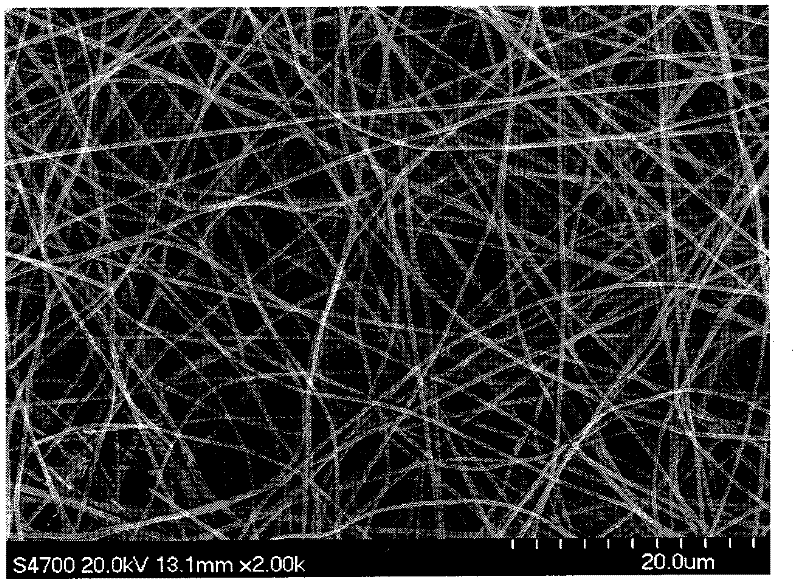
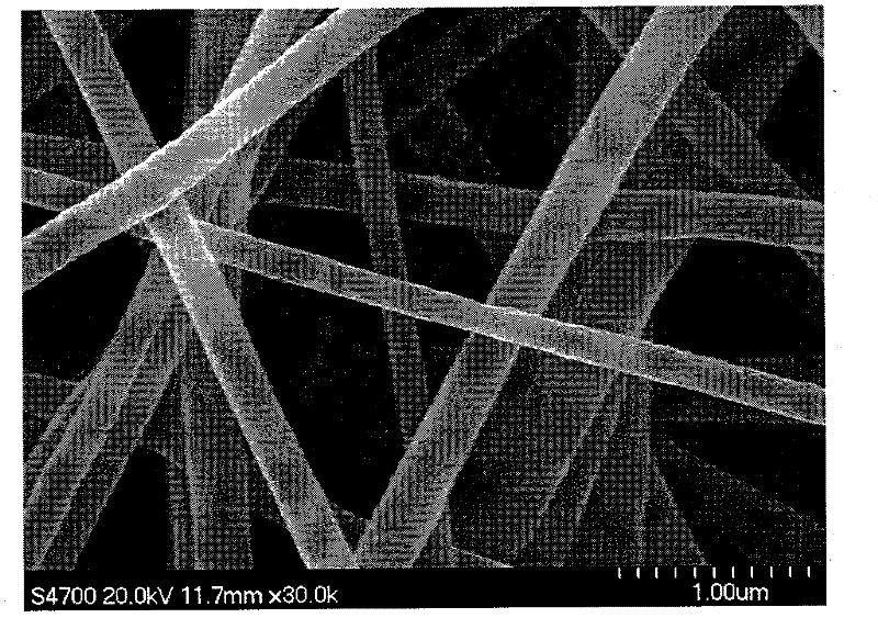
[0033](2) Add the CS electrospinning solution prepared in step (1) into the syringe pump, adjust the voltage to 20kV; the distance from the spinneret to the collecting plate is 12cm; the flow rate of the spinneret spinning solution is 0.5mL / h. The device was activated for electrospinning, and CS nanofibers (such as figure 1 ).

[0034] (3) Cross-link the CS nanofibers obtained in step (2) with glutaraldehyde for 2 hours, soak the cross-linked chitosan nanofibers in the solvent of the pore-forming agent, and selectively remove the pore-forming agent ...

Example Embodiment

[0036] Example 2:

[0037] (1) HA powder of Mw=1,000,000 and PVP (its weight ratio is HA / PVP=5 / 5) are dissolved in deionized water, formic acid and N, in the mixed solvent that N-dimethylformamide forms (deionized Water / formic acid / N, N-dimethylformamide=25 / 50 / 25), be made into the solution of 1wt% by weight percent, then the solution is fully stirred, so that dissolve completely, the solution is placed in ultrasonic for 2 hours to make All the bubbles are separated out to obtain a transparent HA solution.

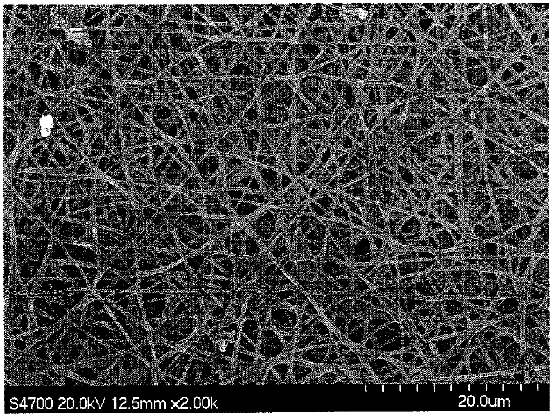
[0038] (2) Add the HA electrospinning solution prepared in step (1) to the syringe pump, adjust the voltage to 15kV; the distance from the spinneret to the collecting plate is 15cm; the flow rate of the spinneret spinning solution is 0.5mL / h; The spinning ambient temperature was 18°C. The device was activated for electrospinning of hyaluronic acid, and HA nanofibers (such as Figure 4 ).

[0039] (3) Cross-linking the hyaluronic acid nanofibers obtained in step (2) wit...

Example Embodiment

[0041] Example 3:

[0042] (1) The mixture of CS powder and PEG (its weight ratio is CS / PEG=9 / 1) is dissolved in the acetic acid solution of 10wt%, is made into the solution that weight percent is 8wt%, then fully stirs solution, so that completely Dissolved, the solution was placed in ultrasonic for 2 hours to make all the bubbles precipitate out to obtain a transparent CS electrospinning solution.

[0043] (2) Add the CS electrospinning solution prepared in step (1) into the syringe pump, adjust the voltage to 25kV; the distance from the spinneret to the collecting plate is 20cm; the flow rate of the spinneret spinning solution is 1mL / h. The device was activated for electrospinning, and CS nanofibers were obtained on the collecting plate.

[0044] (3) The CS nanofibers obtained in step (2) were crosslinked with genipin for 2 hours, and the crosslinked chitosan nanofibers were soaked in the solvent of the pore-forming agent to selectively remove the pore-forming agent and e...


PUM


Abstract

The invention relates to a method for preparing a drug-loaded biodegradable nanofiber medical dressing. The dressing is a nanofiber film, mainly co-spun hyaluronic acid (chitosan) and pore-forming agent to obtain composite nanofibers, using a cross-linking agent to cross-link hyaluronic acid (chitosan), and then selectively The porous nanofibers of hyaluronic acid (chitosan) are obtained by removing the pore-forming agent and cross-linking agent. Finally, the therapeutic drug is encapsulated in the hyaluronic acid (chitosan) nanofibers by using the free combination of polycations and anions. The obtained drug-loaded biodegradable nanofiber medical dressing can absorb the infiltrate well, keep the surrounding area moist, and can release therapeutic drugs continuously for a long time. growth and regeneration, can accelerate wound healing and other functions.

Description

technical field [0001] The invention relates to a method for preparing a drug-loaded biodegradable nanofiber medical dressing. Background technique [0002] Electrospinning is a processing method in which charged polymer solutions or melts are jetted in an electrostatic field to prepare polymer ultrafine fibers. When the applied electric field reaches a certain critical value, the fibers will be ejected from the spinneret At the same time, the solvent gradually evaporates away from the charged fibers, and the dried fibers fall on the collecting plate to form ultra-fine nanofiber non-woven fabrics. The prepared nanofiber non-woven fabric has the advantages of large specific surface area, high porosity, fineness and uniformity of fibers, and large aspect ratio. Generally, the smaller the material system for carrying drugs, the easier it is for the drugs to be absorbed by the body. Therefore, nanofibers have great application possibilities in the sustained release of drugs. ...

Claims


Application Information

Patent Timeline
IPC IPC(8): A61L15/28A61L15/44
Inventor 聂俊马贵平梁鹏总孟准
Owner HANGZHOU INST OF ADVANCED MATERIAL BEIJING UNIV OF CHEM TECH
Why Eureka
Try Eureka