

Glioma-targeting molecule magnetic resonance contrast agent as well as preparation method and application thereof
A molecular magnetic resonance and glioma targeting technology, applied in the field of molecular imaging, can solve the problems of low non-specific binding, cumbersome operation, and large amount of avidin
- Summary
- Abstract
- Description
- Claims
- Application Information
AI Technical Summary
Problems solved by technology
Method used
Image

Examples
Embodiment 1
[0085] 1. Preparation of sterically stable liposomes
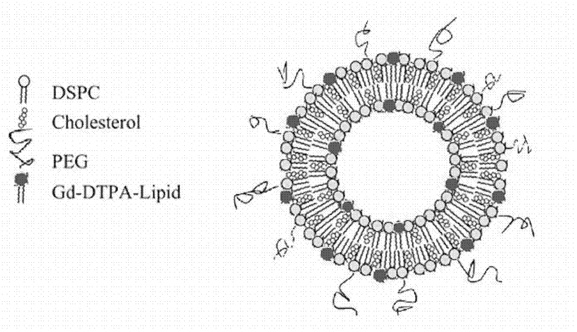
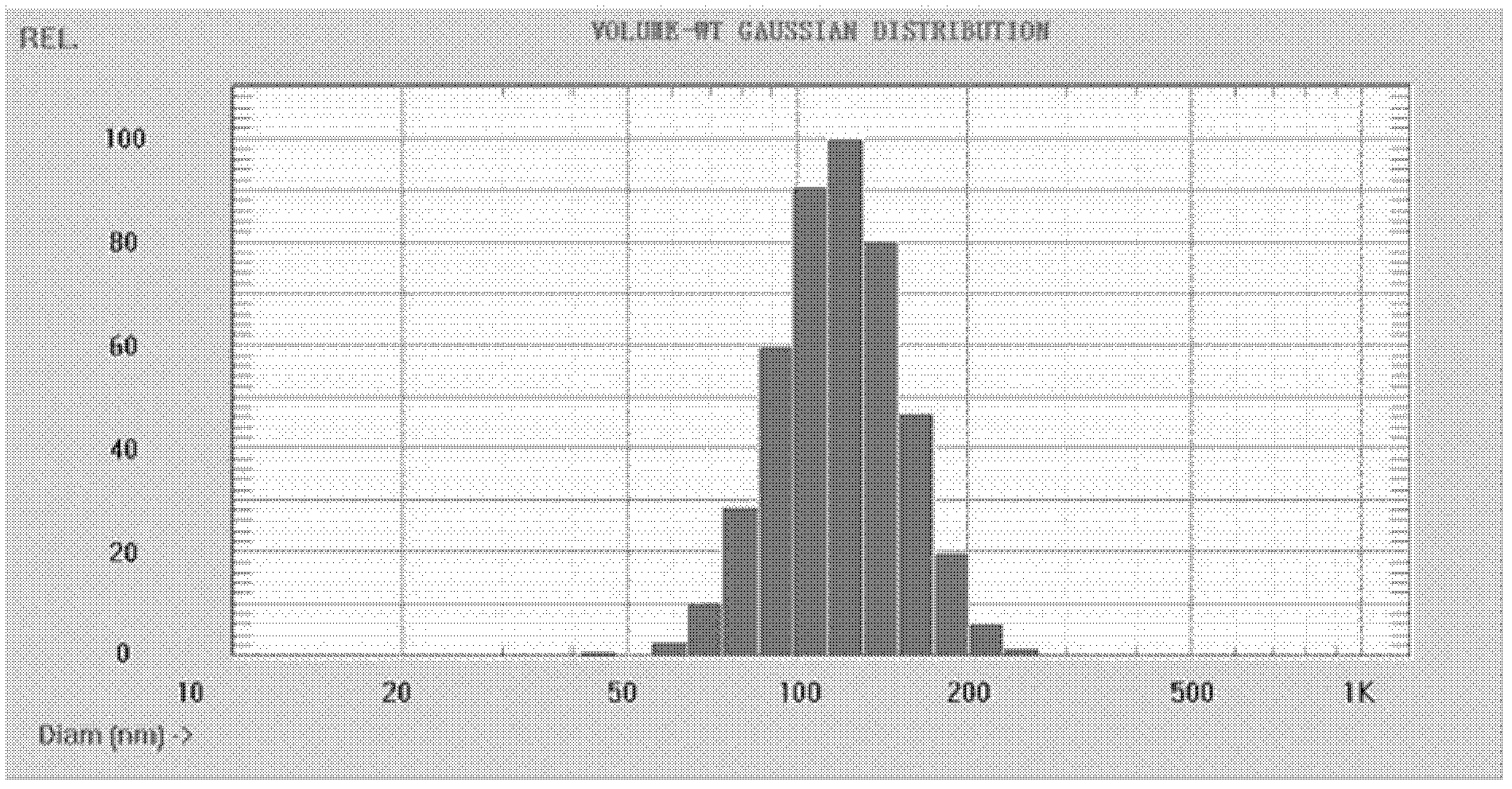
[0086] Liposome membrane material (DSPC / CHOL / mPEG-DSPE / Gd-DTPA-BSA) is dissolved in appropriate amount of anhydrous chloroform and methanol mixed solvent (volume ratio 2: 1) by 36 / 36 / 3 / 25 molar ratio, 40 The solvent was removed by rotary evaporation at ℃, and the lipid film was prepared, and the trace solvent was removed overnight in a vacuum oven at room temperature. Add HEPES buffer (HBS, 20 mM HEPES, 135 mM NaCl, pH=6.5) in a certain proportion (100 μmol lipid: 3 ml), vortex vigorously, hydrate in a water bath at 60 ° C for 2 hours to form a lipid suspension. The suspension was sequentially extruded through a nuclear pore membrane with a pore size of 400 nm, 200 nm, and 100 nm in a high-pressure homogenizer at 60°C for 10 times each to obtain liposomes with a particle size of about 100-120 nm.
[0087] 2. Preparation of PDP-sterically stabilized liposomes (PDP-SLs)
[0088] The preparation method of the PDP-sterically...
Embodiment 2
[0110] Example 2 MR Molecular Imaging Experiment of Endoglin Targets in Rat Glioma Neovascular Endothelial Cells
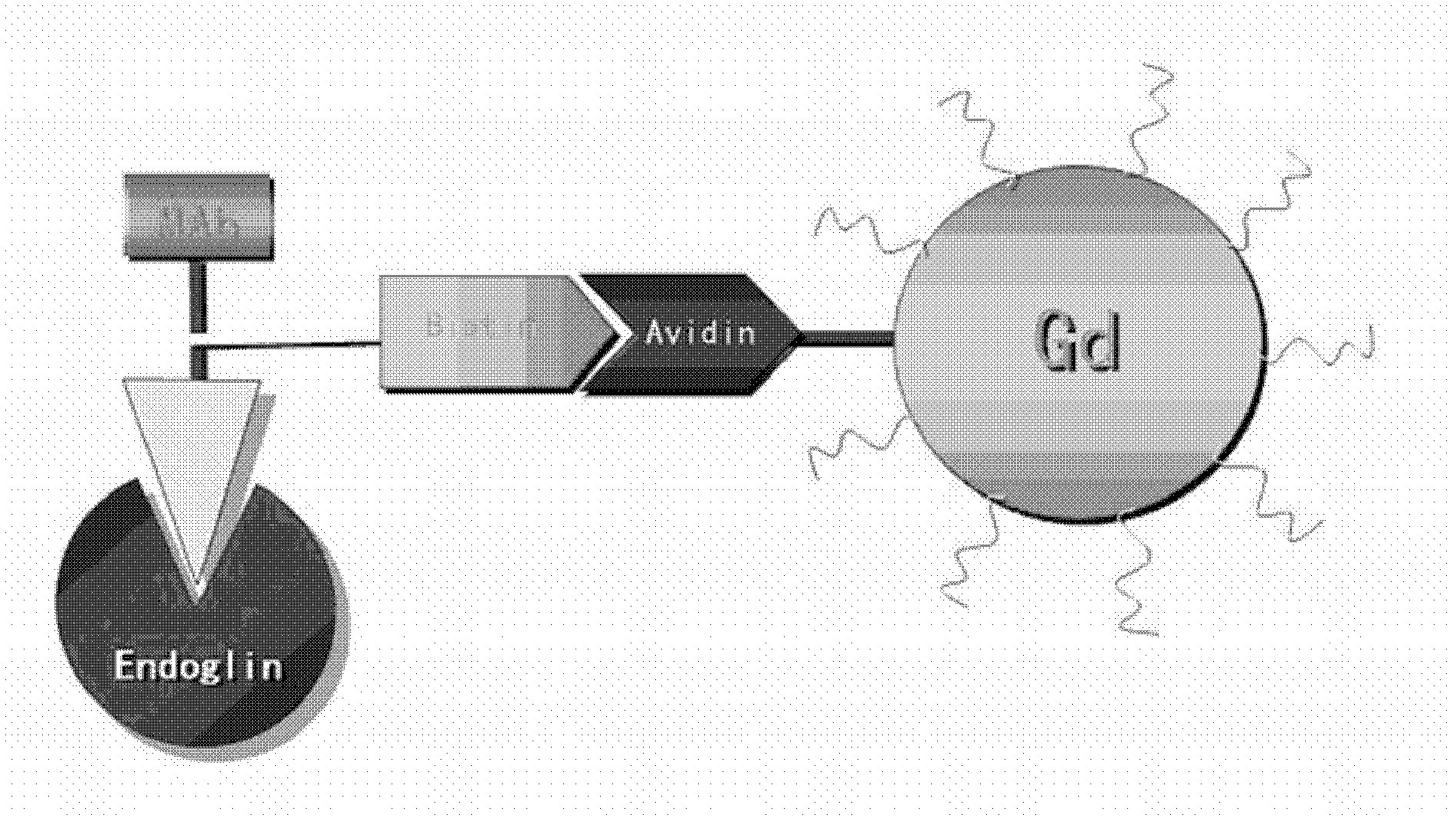
[0111] 25 established rat orthotopic glioma animal models were randomly divided into 5 groups A-E, with 5 rats in each group, and injected gadopentetate dimeglumine (Gd-DTPA) and blank liposome (Gd-DTPA) into the tail vein respectively. -SLs), isotype control IgG liposomes (IgG-SLs), immunoliposome probes combined with Endoglin monoclonal antibody (MAb-SLs) for MR imaging, two-step pre-positioned imaging rats pre-administered biological Streptavidin liposome probes (SAv-SLs) were given after the primed monoclonal antibody (Bio-MAb) for imaging; the differences in tumor enhancement performance in MR T1-weighted enhanced images of rats in each group were compared, and the Time-signal enhancement characteristics of arteries, normal brain tissue, muscle tissue, and tumor tissue; compare the differences in the degree and range of tumor enhancement among the groups, and...
PUM
 Abstract
Description
Claims
Application Information