
CRP monoclonal antibody nanometer latex microsphere composition and preparation process thereof

A monoclonal antibody and latex microsphere technology, applied in the field of medical immunity, can solve the problems of narrow linear range, inability to cover trace CRP, inability to detect CRP, etc., and achieve the effect of small difference, wide application and stable quality.

Active Publication Date: 2013-05-01
深圳伯美生物医药有限公司
View PDF1 Cites 38 Cited by
  • Summary
  • Abstract
  • Description
  • Claims
  • Application Information

AI Technical Summary

Problems solved by technology

CRP detection is roughly divided into two categories according to its concentration detection range: the first type is ordinary CRP detection, the detection range is usually 3-200mg / L, but its linear range cannot cover trace CRP
The second type is high-sensitivity CRP (high-sensitivity CRP, hs-CRP) detection, which is more sensitive than conventional detection methods. It is used to assess the risk of heart disease, but high-sensitivity CRP reagents cannot detect higher concentrations of CRP
These kits have the following deficiencies: First, although polyclonal antibody latex microspheres have higher sensitivity than monoclonal antibody latex microspheres, the linear range is narrower, and it is difficult to meet the requirements of CRP full-range measurement when used alone; secondly, single particle size The microsphere-coupled CRP polyclonal antibody was used to prepare latex microspheres to enhance the immune turbidimetric reagent, and it was impossible to simultaneously measure the CRP content at high and low concentrations.

Method used


Image

  • CRP monoclonal antibody nanometer latex microsphere composition and preparation process thereof
  • CRP monoclonal antibody nanometer latex microsphere composition and preparation process thereof
  • CRP monoclonal antibody nanometer latex microsphere composition and preparation process thereof

Examples


Embodiment 1

[0035] Preparation of CRP monoclonal antibody

[0036] 1 Separation and purification of human serum CRP

[0037] 1.1 Ammonium sulfate precipitation of total protein

[0038] Precipitate the total protein in human serum samples (CRP>100mg / L) with ammonium sulfate with a saturation of 80%, and precipitate overnight at 4°C; the next day, centrifuge at 8000rpm, 4°C for 30min, and wash with basic buffer (20mM Tris-HCl ,pH7.8) to dissolve the precipitate, fully dialyzed to remove ammonium sulfate.

[0039] 1.2 Primary purification of CRP

[0040] Take an appropriate amount of activated DEAE filler (Diethylaminoethyl cellulose) to pack into the column, and equilibrate with the above-mentioned basic buffer. Load the above-mentioned pretreated sample, and carry out gradient elution by changing the ionic strength (the NaCl concentration in the eluent is 80mM, 120mM, and 250mM respectively), collect the eluate respectively, and measure the protein concentration with a UV spectrophotom...

Embodiment 2

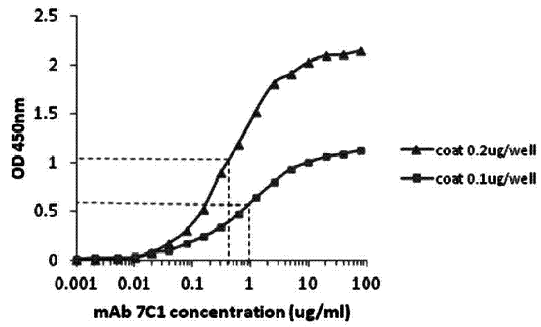
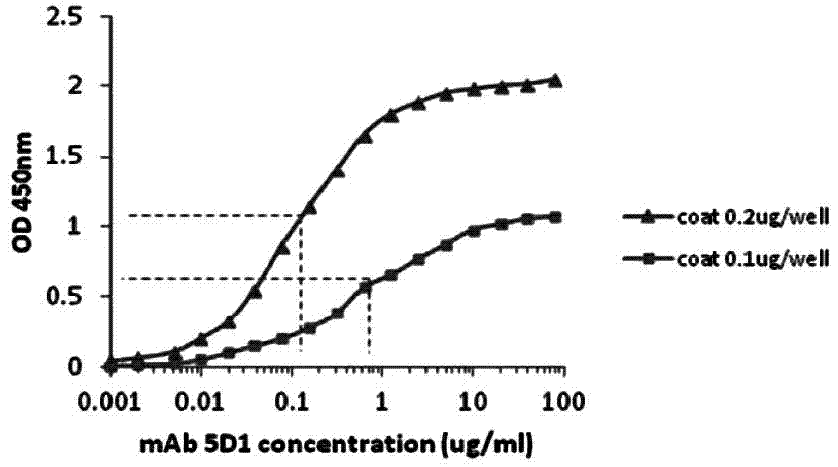
[0071] Determination of Affinity Constant of CRP Monoclonal Antibody by Direct ELISA

[0072] Experimental materials: mouse anti-human CRP monoclonal antibody, from the culture supernatant of CRP monoclonal antibody cell line, filtered through a 0.22 μm filter membrane, affinity purified by Sepharose Protein G, and filtered by Superdex 200 gel, and the isolated group of 200kD-100kD was collected After being concentrated to a certain concentration by ultrafiltration, it was filtered through a 0.22 μm filter membrane and stored in PBS buffer plus 20% (v / v) glycerol at -20°C. The titer determined by indirect ELISA was 2.5×10 6 .

[0073] Experimental procedure: use non-competitive enzyme immunoassay to detect monoclonal antibody affinity constant. The above-mentioned purified human CRP protein was plated and fixed at 0.1 μg and 0.2 μg per reaction well, respectively, using monoclonal antibodies 7C1 and 5D1 as primary antibodies, and horseradish peroxidase (HRP)-coupled rabbit a...

Embodiment 3

[0075] Identification of CRP monoclonal antibodies 7C1 and 5D1 with different epitopes by sandwich ELISA

[0076] Experimental material: see embodiment 2.

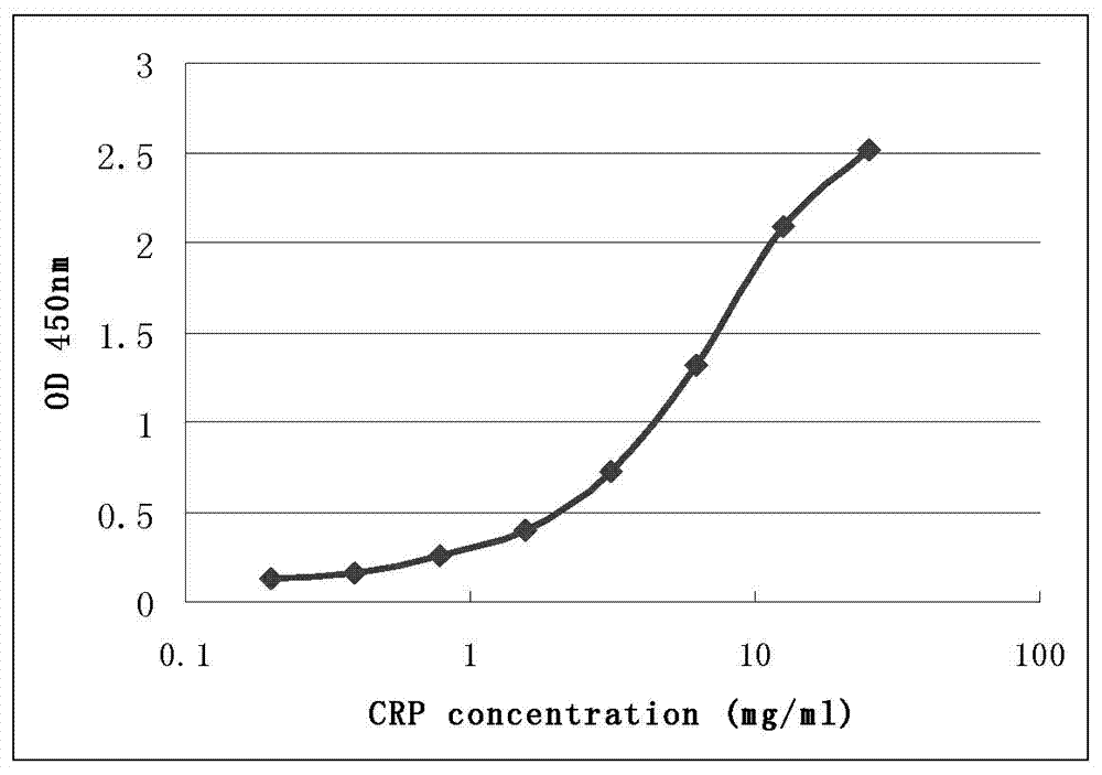
[0077] Experimental procedure: select two complementary monoclonal antibodies 7C1 and 5D1, and detect whether their antigenic epitopes are different. Each reaction well was coated with 0.2 μg monoclonal antibody 7C1 as a capture antibody on the solid phase carrier, and each reaction well was used as a detection antibody with 0.02 μg horseradish peroxidase (HRP)-coupled monoclonal antibody 5D1. Human C-reactive protein was purified and quantified by BCA method, then diluted to different concentrations with PBS buffer, and then detected by sandwich ELISA method. by OD 450nm Reading detection. The test results are shown in Table 1. Take the data in Table 1 as figure 2 . figure 2 Ordinate is OD 450nm The measured value of , the abscissa is the logarithmic value of different CRP antigen concentrations (mg / L).

[0078]...


PUM

PropertyMeasurementUnit
Diameteraaaaaaaaaa
Sensitivityaaaaaaaaaa

Abstract

The present invention discloses a CRP monoclonal antibody, a CRP antibody nanometer latex microsphere composition and a preparation process thereof. According to the CRP antibody nanometer latex microsphere composition, CRP monoclonal antibodies with different epitopes and carboxylated polystyrene microspheres with different particle sizes are subjected to covalent cross-linking to form conjugates, and then the different conjugates are mixed according to a certain ratio to prepare the CRP monoclonal antibody nanometer latex microsphere composition. The CRP monoclonal antibody nanometer latex microsphere composition can be applicable for automatic biochemical analyzers and special protein analyzers, can be provided for full measuring range determination of C-reactive protein concentration in human whole blood and serum, and can be used in differential diagnosis of bacterial infections and viral infections, drug therapy monitoring and cardiovascular disease risk assessments.

Description

technical field [0001] The invention belongs to the field of medical immunity, and relates to the preparation of C-reactive protein (C-reactive protein, CRP) monoclonal antibody (Monoclonal antibody, McAb), the coupling of C-reactive protein monoclonal antibody nano-latex microspheres, and different C-reactive proteins. The combination of protein-antibody nanoemulsion microsphere conjugates also involves the detection application of C-reactive protein. Background technique [0002] C-reactive protein is a protein that binds to pneumococcal cell wall C polysaccharide that can be found in elevated concentrations in the serum of patients with acute inflammation. Human C-reactive protein can exist in at least two forms, one is pentameric CRP (pCRP) formed by non-covalent bonds of five identical subunits, which is the main form of CRP in serum; the other is The monomer (modified / monomeric CRP, mCRP), which is a single subunit, can be formed by the decomposition of pentamer on th...

Claims


Application Information

Patent Timeline
IPC IPC(8): C07K16/18C07K17/08G01N33/68
Inventor 王雷李一凡
Owner 深圳伯美生物医药有限公司
Learn More