Preparation of PEI-coated bimodal contrast agent ferriferrous oxide-gadolinium hydroxide magnetic nanoparticle

A magnetic nanoparticle, dual-modal contrast agent technology, applied in the directions of preparations for in vivo experiments, emulsion delivery, drug delivery, etc.

Inactive Publication Date: 2013-11-27
DONGHUA UNIV +1
View PDF2 Cites 5 Cited by
  • Summary
  • Abstract
  • Description
  • Claims
  • Application Information

AI Technical Summary

Problems solved by technology

[0004] Searching domestic and foreign literatures and patents on the synthesis of dual-mode contrast agent iron ferric oxide-gadolinium hydroxide magnetic nanoparticles wrapped in PEI showed that the Fe synthesiz

Method used


Image

  • Preparation of PEI-coated bimodal contrast agent ferriferrous oxide-gadolinium hydroxide magnetic nanoparticle
  • Preparation of PEI-coated bimodal contrast agent ferriferrous oxide-gadolinium hydroxide magnetic nanoparticle
  • Preparation of PEI-coated bimodal contrast agent ferriferrous oxide-gadolinium hydroxide magnetic nanoparticle

Examples


Embodiment 1

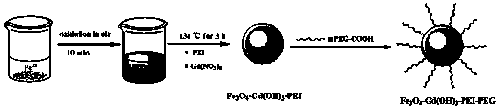
[0047] (1) 1.25g Fe source (FeCl 2 4H 2 O) Put it into a beaker, add 7.75mL of ultrapure water (resistivity greater than 18.2MΩ.cm) to dissolve it. With constant stirring, 6.25 mL of NH 3 ·H 2 O, and continuously stir in the air for 10-15 minutes to ensure that the ferrous iron is oxidized. The reactants were then transferred to the reactor. 2.83g Gd(NO 3 ) 3 Dissolve in 6mL aqueous solution, ultrasonically dissolve, transfer to the reaction kettle, and mix well with the solution in the reaction kettle. At the same time, 0.51g of PEI was dissolved in 3mL of aqueous solution, transferred to the reaction kettle, and fully mixed with the solution in the reaction kettle. The mixture was reacted at 134-140°C for about 3-3.5 hours. After the reaction, cool down to room temperature naturally, disperse the obtained black precipitate in ultrapure water, magnetically separate, disperse again, and magnetically separate, repeat this five times of pure water washing to remove exces...

Embodiment 2

[0053] The above-prepared Fe 3 o 4 -Gd(OH) 3 -PEI-PEG nanomaterials were measured by the ICP-AES test method to measure the content of Fe and Gd elements in the solution, and then use EP tubes to prepare 2 mL of aqueous solutions with different Fe concentrations and Gd concentrations, and pass T 1 and T 2 Magnetic Resonance Imaging Study Materials T 1 and T 2 relaxation effect. Figure 5 and 6 Respectively Fe 3 o 4 -Gd(OH) 3 - Gray-white graph and linear fitting graph of PEI-PEG nanoparticles as a function of Gd or Fe concentration, from Figure 5 a The gray and white figure shows that with the increase of Gd concentration, Fe 3 o 4 -Gd(OH) 3 - Signal enhancement of PEI-PEG nanoparticles, which has a good r 1 Relaxation rate (5.63mM -1 the s -1 );From Figure 6 a The gray and white figure shows that with the increase of Fe concentration, Fe 3 o 4 -Gd(OH) 3 -The signal of PEI-PEG nanoparticles is weakened, as can be seen from the linear relationship diagram o...

Embodiment 3

[0055] Using KB cells as model cells, the dual-modal contrast agent Fe synthesized by MTT colorimetry was used to measure the viability of KB cells (a cell line of human epithelial cancer) 3 o 4 -Gd(OH) 3 - Cytotoxicity of PEI-PEG nanomaterials. KB cells and Fe 3 o 4 -Gd(OH) 3 - PEI-PEG (concentrations of 10, 25, 50 and 100 μg / mL) were co-cultured at 37°C for 24 hours. Then, the absorbance value was measured at 570nm after MTT treatment, and the cell proliferation and viability were calculated according to this value. From Figure 7 It can be seen that compared with the control group, in the range of 0 to 100 μg / mL, the Fe 3 o 4 -Gd(OH) 3 -The cells treated with PEI-PEG all had good cell viability. The effect of different concentrations of materials on cell proliferation was analyzed by one-way analysis of variance. Compared with the control group (buffer PBS, pH7.4), Fe 3 o 4 -Gd(OH) 3 -PEI-PEG has no significant difference in the viability of KB cells in the tes...


PUM

PropertyMeasurementUnit
Diameteraaaaaaaaaa

Abstract

The invention relates to a preparation method for a PEI-coated bimodal contrast agent, i.e., a ferriferrous oxide-gadolinium hydroxide magnetic nanoparticle. The preparation method comprises the following steps: (1) dissolving an Fe source in ultrapure water, then adding NH3.H2O, carrying out stirring in the air to realize oxidation, then adding an aqueous solution of polyethylene imine (PEI) and an aqueous solution of Gd(NO3)3 and carrying out natural cooling to room temperature after completion of a reaction so as to obtain Fe3O4-Gd(OH)3-PEI; and (2) subjecting Fe3O4-Gd(OH)3-PEI and methoxypolyethylene glycol carboxylic acid to pegylation. The preparation method provided by the invention has the advantages of simple process, mild reaction conditions and easy operation and separation; the prepared ferriferrous oxide-gadolinium hydroxide magnetic nanoparticle (Fe3O4-Gd(OH)3-PEI-PEG) has good biocompatibility, good relaxation effects at T1 and T2 and a latent application value in the field of MRI imaging diagnosis.

Description

technical field [0001] The invention belongs to the field of preparation of modal magnetic nanoparticles, and in particular relates to the preparation of dual-mode contrast agent iron ferric oxide-gadolinium hydroxide magnetic nanoparticles wrapped in PEI. Background technique [0002] At present, cancer has become one of the deadliest "killers" of human beings. Due to the local invasion and distant metastasis of malignant tumors, the treatment effect and cost of cancer are closely related to the period of disease discovery. Therefore, early detection of tumors is the key to the treatment of cancer. Molecular imaging can detect the molecular biological characteristics of the disease, such as precancerous molecular changes, gene changes and other tumor markers, before clinical symptoms appear in the early stage of the tumor, so as to realize early detection and treatment of tumors. In recent years, with the development of nanotechnology, the emerging field of nanomedicine ha...

Claims


Application Information

Patent Timeline
IPC IPC(8): A61K49/12A61K49/18
Inventor 史向阳蔡红东温诗辉沈明武安潇
Owner DONGHUA UNIV
Why Eureka
Try Eureka