
Ultra wide band stimulated raman spectroscopy microscopic imaging system simple and convenient to use

A microscopic imaging and stimulated Raman technology, which is applied in Raman scattering, spectrometry/spectrophotometry/monochromator, material excitation analysis, etc., can solve problems such as expensive, complex system, and bulky , to achieve the effect of reducing construction cost, convenient operation and reducing volume

Active Publication Date: 2014-01-01
SOUTH CHINA NORMAL UNIVERSITY
View PDF4 Cites 19 Cited by
  • Summary
  • Abstract
  • Description
  • Claims
  • Application Information

AI Technical Summary

Problems solved by technology

[0004] In a large number of studies in the past, the stimulated Raman microscopy system used two titanium sapphire lasers or a titanium sapphire laser and an optical parametric oscillator as the light sources of the two beams respectively. This dual light source system is expensive. Huge volume, complex system, cumbersome pulse tuning and synchronization, and it is difficult to achieve continuously adjustable ultra-broadband spectral imaging of samples in different Raman spectral ranges, even if some researchers can use some methods to achieve certain Raman spectral imaging. Continuous imaging of the spectral range, but its imaging spectral range is also very limited

Method used


Image

  • Ultra wide band stimulated raman spectroscopy microscopic imaging system simple and convenient to use
  • Ultra wide band stimulated raman spectroscopy microscopic imaging system simple and convenient to use
  • Ultra wide band stimulated raman spectroscopy microscopic imaging system simple and convenient to use

Examples


Embodiment 1

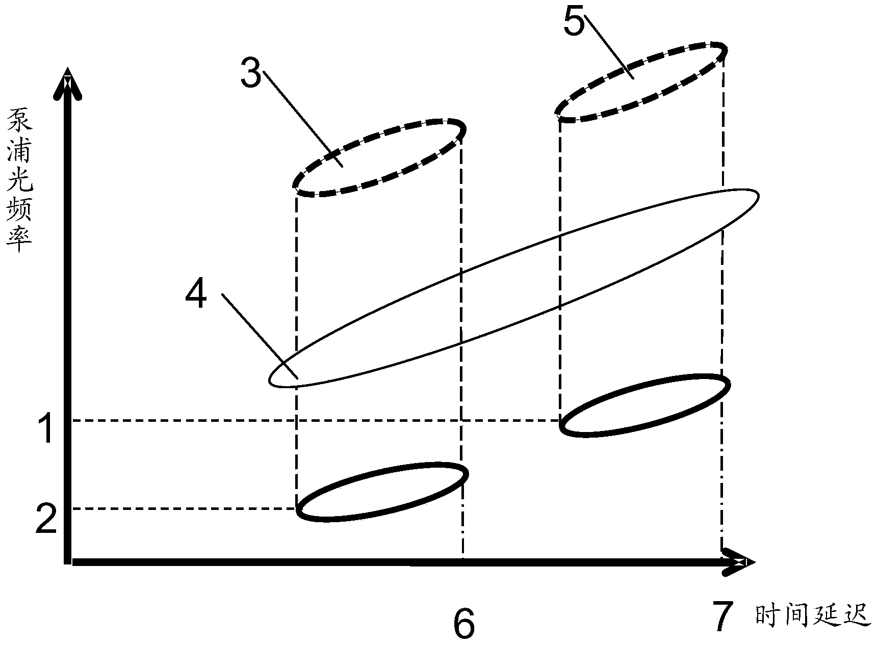
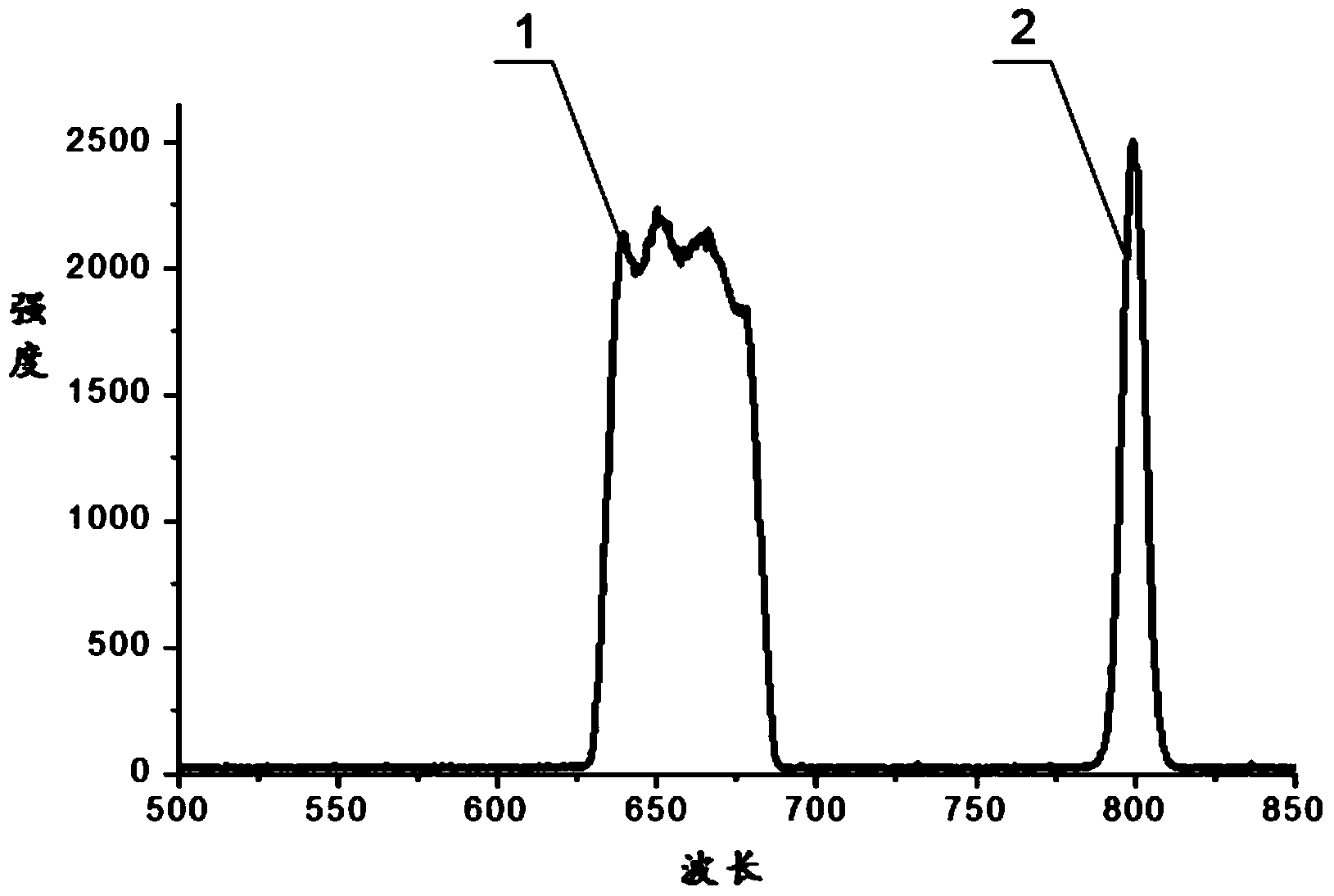
[0022] The supercontinuum generated by the expansion of a single pulse by various nonlinear effects in photonic crystal fibers is a new type of light source. It has the characteristics of high output power, flat broadband spectrum, and high spatial coherence. It can greatly Improve the signal-to-noise ratio and widen the spectral measurement range, and have a wide range of applications in biological imaging, optical fiber attenuation measurement, interferometer, optical coherence photography, optical frequency comb, etc. The supercontinuum has chirp characteristics, and there is a time difference for different wavelengths of a single pulse. Therefore, if the pulsed laser and the different components in the broadband supercontinuum can be synchronized in space and time, it is expected to realize convenient and adjustable Ultra-broadband stimulated Raman continuum imaging, for example, if a relatively common section of the supercontinuum from 600 nm to 790 nm is used as pump ligh...

Embodiment 2

[0028] Present embodiment except following feature other structures are with embodiment 1:

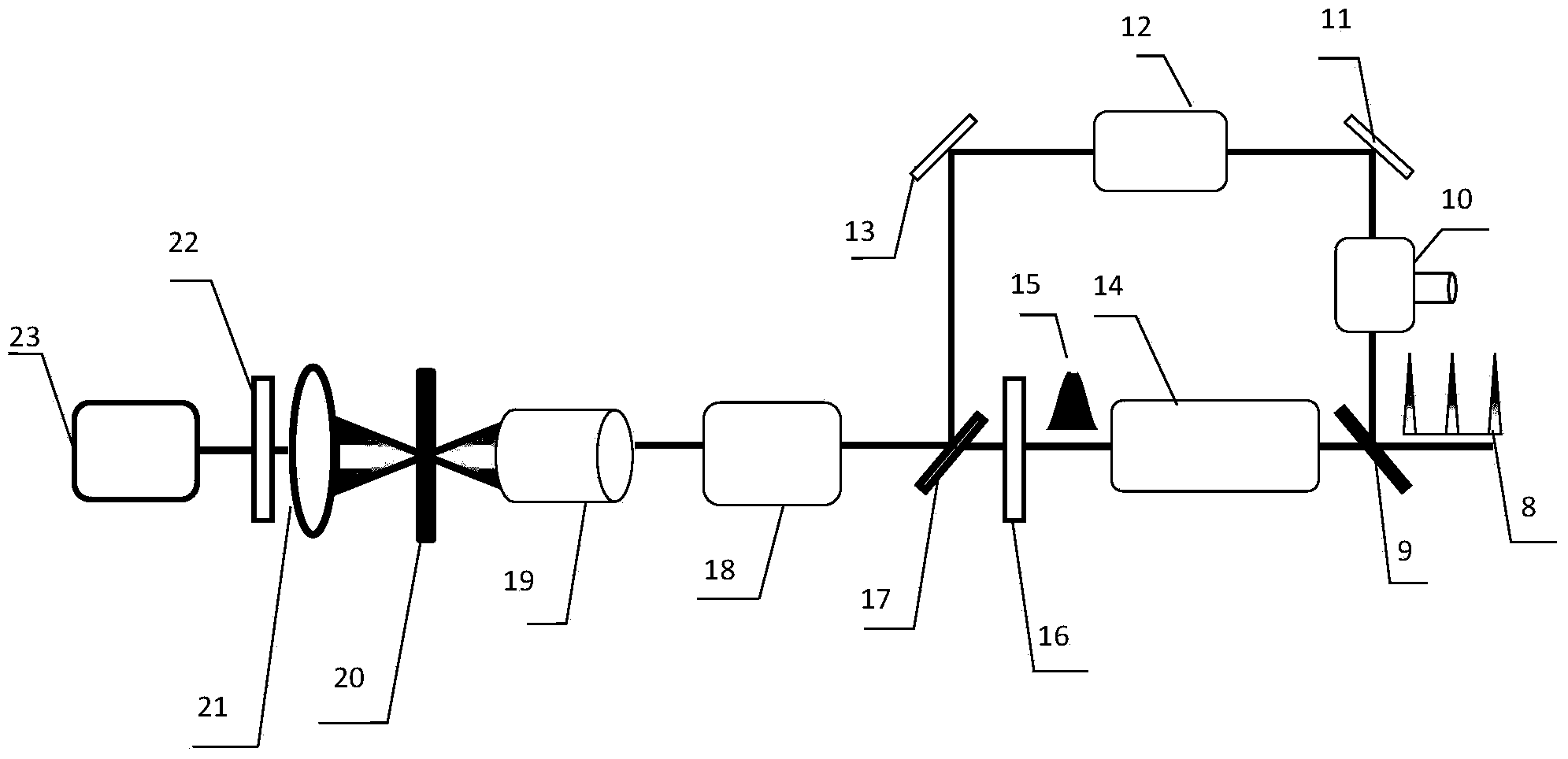
[0029] Such as Figure 4 As shown, the ultra-broadband stimulated Raman spectroscopy microscopic imaging system of this embodiment includes an ultrashort pulse laser (titanium sapphire laser in this embodiment), a polarizing beam splitter 9, a retroreflective mirror system 10, and a light intensity modulator 12 (this embodiment is a chopper), photonic crystal fiber 14, bandpass filter 16, dichroic mirror 17, also includes scanning unit 18, second beam splitter 218, photodetector 220, first objective lens 221 , the second objective lens 223, optical filter 224, balance detector 225, lock-in amplifier 226, first beam splitter 219, in addition, Figure 4 The middle marks 11, 213, 214, 215, 216, 217 are reflecting mirrors.

[0030] The working process is: the 800nm ​​pulse width about 200 femtosecond pulse laser beam 8 produced by the titanium sapphire laser is divided into two beams of ...


PUM


Abstract

The invention discloses an ultra wide band stimulated raman spectroscopy microscopic imaging system simple and convenient to use. The ultra wide band stimulated raman spectroscopy microscopic imaging system is obtained through the method including the steps that ultra-short pulse lasers generated by an ultra-short pulse laser are divided into two beams through a spectroscope, one of the beams is strokes light which sequentially passes through an adjustable retroreflection mirror system with a rotary knob, and a light intensity modulator, the other beam generates a super-continuum spectrum through a photonic crystal fiber, and a part of the wave band of the super-continuum spectrum is selected through a band-pass filter to be used as pump light, time relay of two pulses is rapidly adjusted through the retroreflection mirror system after the two beams are converged through a dichroscope, the components, with different wavelengths, of the strokes light and the components, with different wavelengths, of the pump light are overlapped, sequentially pass through a scanning unit, an objective lens, an optical filter and a photoelectric detector, and are processed through a lock-in amplifier and imaged on a computer, the continuous adjustable ultra wide band stimulated raman biological microscope spectral imaging is finally achieved, and the ultra wide band spectral component continuous adjustable stimulated raman spectrum detection imaging which has great research value in the bioscience field can be specifically achieved.

Description

technical field [0001] The invention relates to the fields of optical microscopic technology, optical fiber technology and biological detection technology, in particular to a simple ultra-broadband stimulated Raman spectrum microscopic imaging system. Background technique [0002] In optical bioimaging systems, probe-labeled fluorescence microscopy, especially confocal fluorescence microscopy based on fluorescent single-molecule processes, has been widely used due to its advantages of high resolution, high sensitivity, and high magnification. However, since some biomolecules have no obvious photoluminescent properties or labeled probes are easy to cause biological damage, new label-free, high-specificity, high-contrast bioimaging methods, such as stimulated Raman scattering imaging, Coherent anti-Stokes Raman scattering imaging, stimulated fluorescence imaging, etc. [0003] Among them, the stimulated Raman scattering microscopic imaging is irradiated by two beams with a sp...

Claims


Application Information

Patent Timeline
IPC IPC(8): G01J3/44G01N21/65
Inventor 詹求强赵宇翔
Owner SOUTH CHINA NORMAL UNIVERSITY
Features
  • Generate Ideas
  • Intellectual Property
  • Life Sciences
  • Materials
  • Tech Scout
  • Unparalleled Data Quality
  • Higher Quality Content
  • 60% Fewer Hallucinations
Learn More