Gene and drug co-transported PLGA ultrasonic nano bubbles as well as preparation method and application thereof

A technology of PLGA and nanobubbles, applied in gene therapy, powder delivery, freeze-drying delivery, etc., can solve the limitations of diagnosis of extravascular diseases, the large particle size of microbubble-level ultrasound contrast agents, and the inability to cross the "intima barrier". Achieve good drug loading and encapsulation efficiency, low equipment requirements, and long storage time

Inactive Publication Date: 2014-12-24
UNIV OF ELECTRONICS SCI & TECH OF CHINA
View PDF1 Cites 13 Cited by
  • Summary
  • Abstract
  • Description
  • Claims
  • Application Information

AI Technical Summary

Problems solved by technology

[0003] The commonly used microbubble grade ultrasound contrast agent has a large particle size and cannot cross the "intima barrier", and only a small amount of the drug it carries enters into the tumor cells after intravascular release, which limits the diagnosis of many extravascular diseases

Method used


Image

  • Gene and drug co-transported PLGA ultrasonic nano bubbles as well as preparation method and application thereof
  • Gene and drug co-transported PLGA ultrasonic nano bubbles as well as preparation method and application thereof
  • Gene and drug co-transported PLGA ultrasonic nano bubbles as well as preparation method and application thereof

Examples


Embodiment 1

[0030] Example 1: Synthesis of PLGA nanobubbles for co-delivery of genes and drugs

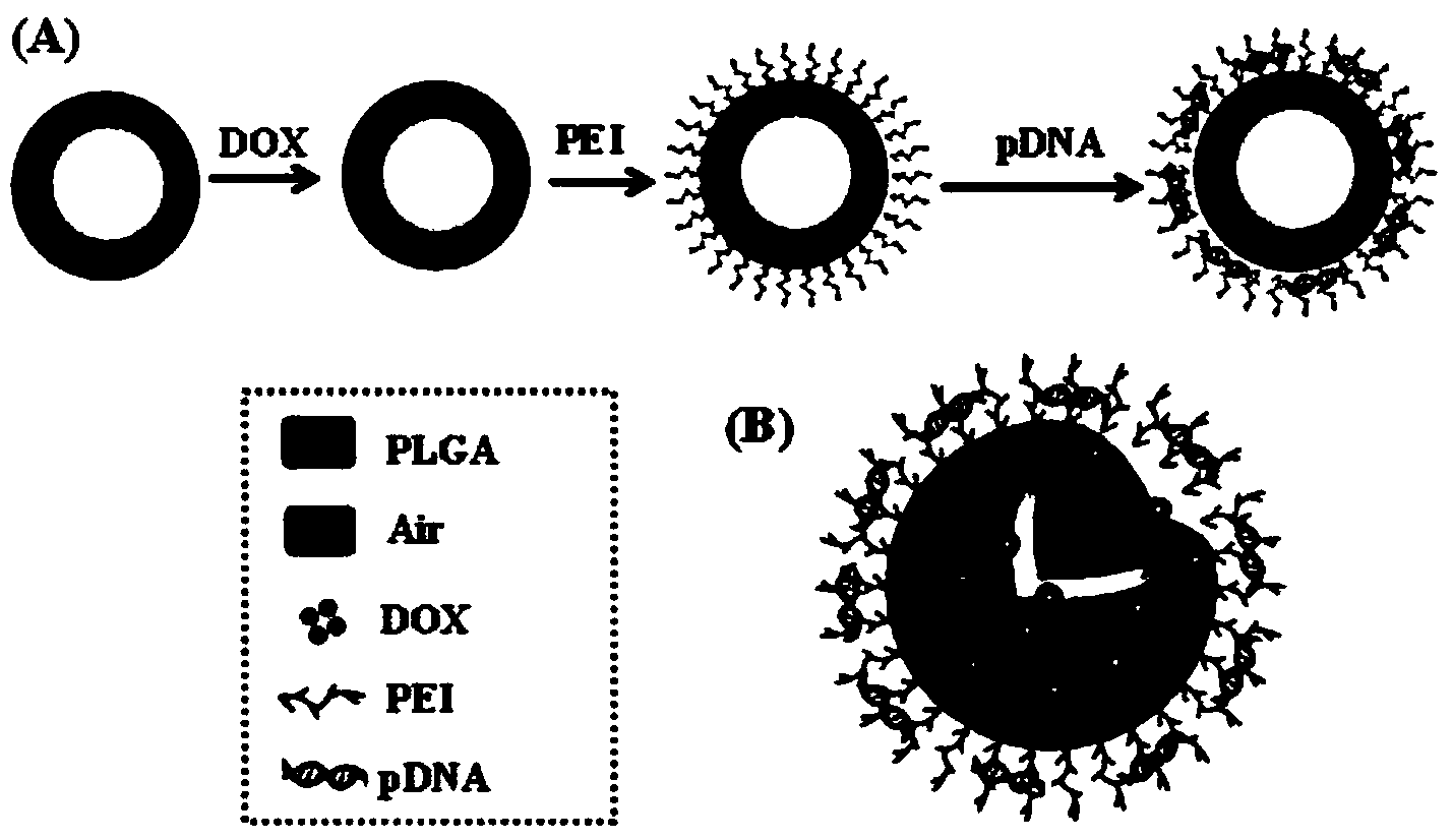
[0031] ①The nanobubble membrane is made of PLGA material, and the anti-tumor drug DOX and air are loaded inside. The surface of the nanobubble is connected with PEI, and the plasmid shRNA that reverses the multidrug resistance gene (MDR-1) is adsorbed. The synthetic schematic diagram and three-dimensional structure diagram are shown in figure 1 shown.

[0032] ② Control the particle size of nanobubbles, within the range of 200-500nm, they are uniform spherical and well dispersed in aqueous solution.

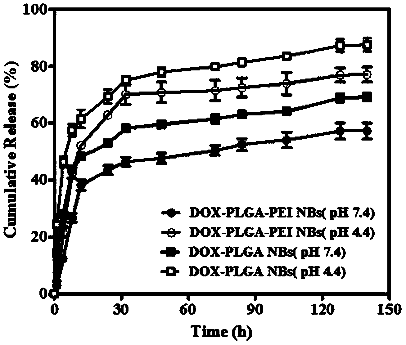
[0033] ③By controlling the particle size of the nanobubbles, they can passively target the tumor site through the EPR effect, which can not only be used for tumor imaging; at the same time, the carried gene is first released under intracellular acidic conditions to silence the multidrug resistance gene MDR-1 expression, reverse tumor drug resistance; at the same time release the carried drug DO...

Embodiment 2

[0034] Example 2: Preparation of drug-loaded PLGA ultrasonic nanobubbles

[0035] ①Weigh 0.2g PLGA with an electronic analytical balance and place it in 5ml dichloromethane (CH 2 Cl 2 ) glass bottle, completely dissolved.

[0036] ②Add 1.0ml of DOX solution to the dissolved PLGA dichloromethane solution, use ultrasonic breaker to form W / O primary emulsion after ultrasonic emulsification at 20% ultrasonic amplitude for 2 minutes.

[0037] ③ Quickly pour the W / O primary emulsion into 30ml of 1% PVA solution, and continue ultrasonic emulsification for 1 minute to form a W / O / W complex emulsion.

[0038] ④ After adding 1.5ml of isopropanol solution dropwise into the complex emulsion, stir with a magnetic stirrer for 4 hours at room temperature.

[0039] ⑤ Divide the above liquid evenly into centrifuge tubes, centrifuge for 10 minutes, discard the supernatant, and collect the sediment. Add an appropriate amount of double-distilled water again, centrifuge, discard the supernatant...

Embodiment 3

[0043] Example 3: Preparation of a PLGA ultrasonic nanobubble for co-delivery of genes and drugs

[0044] ① Prepare DOX-PLGA NBs according to the method in Example 2.

[0045]②Weigh 15mg of DOX-PLGA NBs and dissolve in 1.5ml double distilled water solution, add 400μl EDC solution, and react for 1h.

[0046] ③ Add PEI solution according to the mass ratio of nanobubbles and PEI at 20:1, place in a constant temperature shaker, and react at 37°C for 12h.

[0047] ④ Place the above reaction solution in a centrifuge tube, centrifuge, discard the supernatant, add double distilled water to wash, centrifuge, discard the supernatant. Repeat 5 times in total.

[0048] ⑤After freeze-drying, DOX-PLGA-PEI NBs freeze-dried powder was obtained, and stored at 4°C for future use.

[0049] ⑥ Mix the synthesized DOX-PLGA-PEI NBs and pDNA at a mass ratio of 50:1 before the experiment, and let it stand at room temperature for more than 30 minutes before use.

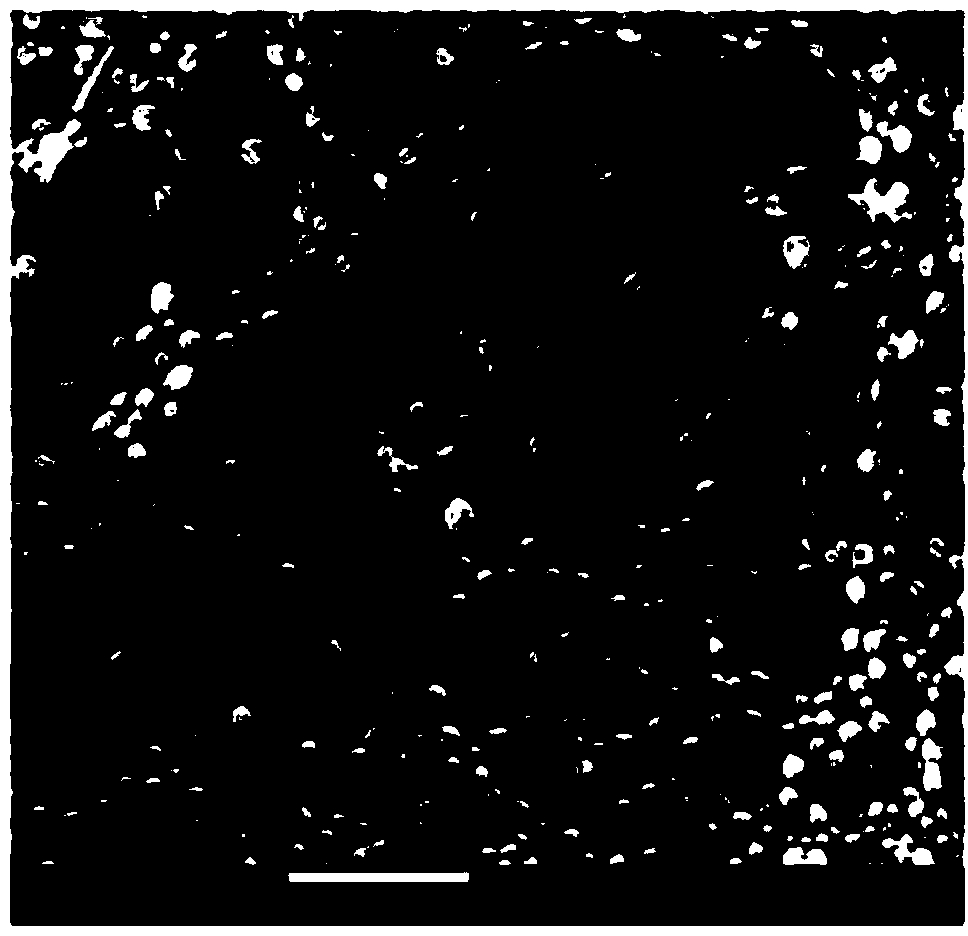
[0050] The electron microscope pic...


PUM

PropertyMeasurementUnit
particle diameteraaaaaaaaaa

Abstract

The invention relates to PLGA ultrasonic nano bubbles integrating ultrasonic imaging, drug delivery and gene therapy into a whole and a preparation method thereof. The preparation method comprises the following steps: preparing drug-loading nano bubbles loaded with a chemotherapy drug DOX, further modifying PLGA nano bubbles through PEI, adsorbing shRNA plasmids of MDR-1, thereby forming the composite nano bubbles. The nano bubbles have high drug loading rate and encapsulation efficiency, drug release has pH responsiveness, the loaded genes can be protected, expression of drug resistance genes MDR-1 is effectively silenced, the ultrasonic imaging capacity can be exerted, and the drugs and genes can be simultaneously and efficiently delivered to tumor locations to exert a synergistic effect.

Description

technical field [0001] The invention belongs to the field of nanomaterial preparation, and in particular relates to a PLGA ultrasonic nanobubble for co-delivery of genes and drugs, a preparation method and application thereof. Background technique [0002] Ultrasound imaging has been widely used in the clinical diagnosis of diseases due to its advantages of real-time non-invasive, high sensitivity, and low cost, and has become one of the main detection methods of modern medical imaging. The emergence and continuous development of ultrasound contrast agents have greatly enhanced the resolution of ultrasound imaging and improved the contrast. Studies in recent years have shown that ultrasound contrast agents not only have good application value in molecular imaging and thrombosis therapy, but also have achieved great success in in vitro targeted delivery and release of drugs or genes, which suggests that we can combine ultrasound Molecular imaging of contrast agents and targe...

Claims


Application Information

Patent Timeline
Patent Type & Authority Applications(China)
IPC IPC(8): A61K49/22A61K48/00A61K47/34A61K9/19A61P35/00A61K31/704
Inventor 刘贻尧杨红邓力蔚
Owner UNIV OF ELECTRONICS SCI & TECH OF CHINA
Why Eureka
Try Eureka