Experimental apparatus for in-vitro circulating perfusion and digestion of liver tissues

An extracorporeal circulation and experimental instrument technology, applied in biochemical instruments, bioreactors/fermenters for specific purposes, sterilization methods, etc., can solve problems such as experimental failure, low culture success rate, loss of digestive enzymes, etc. Experimental cycle, simple experimental operation, and the effect of reducing difficulty

Inactive Publication Date: 2015-01-28
GUIYANG MEDICAL UNIVERSITY
View PDF5 Cites 9 Cited by
  • Summary
  • Abstract
  • Description
  • Claims
  • Application Information

AI Technical Summary

Problems solved by technology

However, this in situ circulatory digestion method has the following disadvantages: 1. The surgical operation in the experiment is difficult and difficult to master; 2. During the circulatory perfusion process, part of the enzyme solution will leak through the liver capsule and cannot be refluxed through the vena cava catheter; and There are collateral circulation blood vessels between the liver and the diaphragm, and some of them are thin. It is impossible to completely ligate all the collateral vessels. During the circulation process, the enzyme solution will also flow away through the unligated collateral vessels. Therefore, this circulation method must There is a large amount of expensive digestive enzyme loss; 3. After in situ perfusion digestion, the liver capsule softens and is easy to rupture. Once the liver capsule ruptures, it will easily lead to cell loss or contamination, which will lead to the failure of the experiment
This method reduces some operating steps and appropriately reduces the difficulty of the operation, but it still needs to be intubated into the inferior vena cava, and there may be more problems with the loss of digestive enzymes during the circulatory perfusion process. At the same time, the digested liver is removed from the animal. When removed, there are still fatal defects such as cell loss and easy contamination caused by rupture of the liver capsule
[0003] The patent application with the application number "200410084625.7" discloses a "preparation method of pig and human hepatocytes for bioartificial liver". Although it discloses that hepatocytes can be separated by extracorporeal circulation, the application only describes the The method of hepatocytes (including the conditions, reagents, time, steps, etc. in the separation method) does not disclose any content about the extracorporeal circulation perfusion device, so it cannot be implemented in actual application;
[0004] The patent application with the application number "01210227719.X" discloses "a heavy metal adsorbent for extracorporeal perfusion and its preparation method". Although it also involves "in vitro perfusion", it only refers to whole blood extracorporeal perfusion. It is used to adsorb heavy metal ions in blood, so it is not suitable for the separation of various cells in liver tissue and related experimental research on the biological characteristics of various cells in liver tissue
[0005] The patent application with the application number "201410142209.1" discloses a "organ decellularization circulation perfusion regeneration culture flask", although it also involves circulation perfusion, the device is mainly used to remove cells while retaining organ scaffolds (organ scaffolds) during decellularization. It is mainly some collagen tissue. In the experiment, the cells on the organ were digested and detached, leaving a scaffold), and the whole decellularization process took 3-5 days, so these cells were basically inactivated or severely damaged after detachment. The success rate of culture is also extremely low, so this culture flask is not suitable for isolating various cells in liver tissue and conducting relevant experimental research on the biological characteristics of various cells in liver tissue
Therefore, there is an urgent need for an extracorporeal circulation perfusion experimental instrument to realize the separation of various cells in the liver tissue for related experimental research on the biological characteristics of various cells in the liver tissue. The experimental instrument that solves the above problems is disclosed

Method used


Image

  • Experimental apparatus for in-vitro circulating perfusion and digestion of liver tissues

Examples


Embodiment Construction

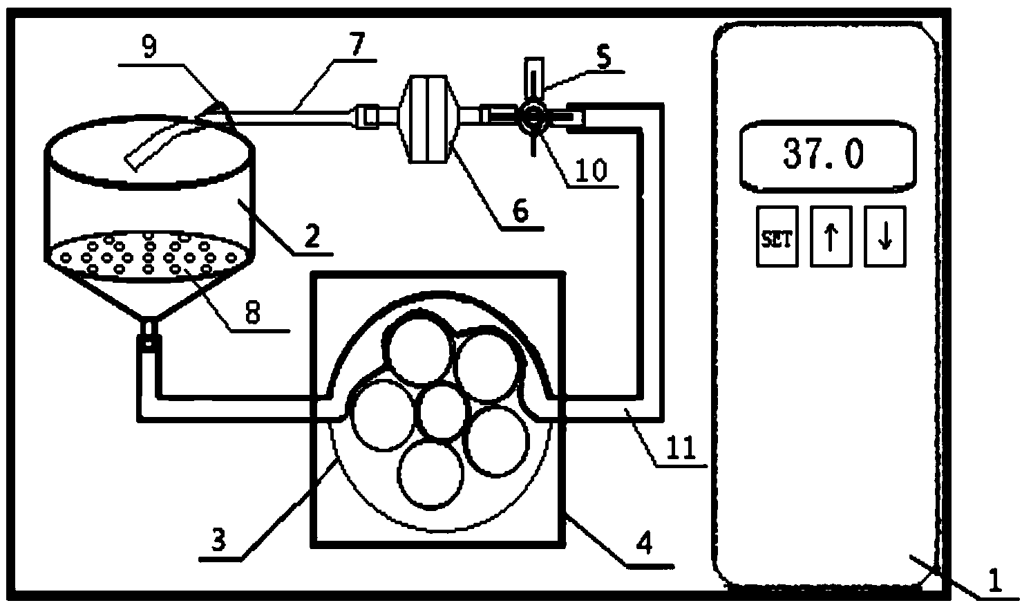
[0026] Embodiments of the present invention: an experimental instrument for extracorporeal circulation perfusion digestion of liver tissue, such as figure 1As shown, it includes an incubator 1 and a perfusion circuit arranged in the incubator 1. The perfusion circuit includes: a container for holding liver tissue 2 connected in sequence, a peristaltic pump device 4, a three-way pipe 5, a filter device 6 and Sterile pipeline 7; the peristaltic pump device 4 includes a peristaltic pump 3 and a flexible pipeline 11 corresponding to the peristaltic pump 3, the flexible pipeline 11 bypasses the corresponding peristaltic pump 3 and the two ends are respectively connected to the liver tissue container 2 and the three Through pipe 5 communicates. The temperature of the thermostat 1 is 37°C. The bottom of the container 2 for holding liver tissue is funnel-shaped, and a partition 8 with holes is arranged inside it. A hook 9 is provided at the top of the container 2 for holding liver t...


PUM


Abstract

The invention discloses an experimental apparatus for in-vitro circulating perfusion and digestion of liver tissues. The experimental apparatus comprises an incubator 1 and a perfusion loop arranged in the incubator 1, wherein the perfusion loop comprises a liver-tissue container 2, a peristaltic pump device 4, a three-way pipe 5, a filtering device 6 and an aseptic pipeline 7 which are connected in sequence; the peristaltic pump device 4 comprises a peristaltic pump 3 and a flexible pipeline 11 corresponding to the peristaltic pump 3; the flexible pipeline 11 bypasses the corresponding peristaltic pump 3 and the two ends of the flexible pipeline 11 are respectively communicated with the liver-tissue container 2 and the three-way pipe 5. The experimental apparatus disclosed by the invention can be applied to separation of various cells in the liver tissues, can digest collagenous tissues in the liver tissues by a circulating digestion mode to enable the cells to be dispersed, and collect the cells for the following culture and realizes related experimental research on the biological characteristics of various cells in the liver tissues; since the cells are separated out in shorter time, the activity of the separated-out cells is higher and the success ratio of anaphase culture is increased.

Description

technical field [0001] The invention relates to an experimental instrument, in particular to an experimental instrument for extracorporeal circulation perfusion digestion of liver tissue. Background technique [0002] In the experimental research on liver-related diseases, it is an essential experimental method to isolate and culture hepatocytes, Kupffer cells, hepatic stellate cells and other cells in liver tissues from experimental animals or even human liver tissues. At present, the isolation of these cells from experimental animals often adopts the method of in situ circulation (in vivo circulation) digestion. The specific method is: after the experimental animal is anesthetized, the abdomen is disinfected, the abdominal cavity is opened, the portal vein and inferior vena cava are exposed, and the portal vein is intubated. , infuse balanced salt solution (perfusate) through the portal vein cannula, and cut the inferior vena cava to let blood out. After the blood in the l...

Claims


Application Information

Patent Timeline
IPC IPC(8): C12M1/00C12M1/12
CPCC12M45/09C12M29/02
Inventor 张金娟杨勤肖俊陈腾祥郭晓婷韩冰谢汝佳
Owner GUIYANG MEDICAL UNIVERSITY
Why Eureka
Try Eureka