Preparation method for TPGS medicine carrying lipidosome-natural material composite nano-fiber bracket
A technology of composite nanofibers and natural materials, which is applied in the field of drug-loaded liposomes composited with natural material nanofiber scaffolds, can solve the problems of limiting clinical application and industrial production, low drug loading rate, thermodynamic instability, etc., and achieve drug loading And the effects of high transfection efficiency, good biocompatibility, and good application prospects
- Summary
- Abstract
- Description
- Claims
- Application Information
AI Technical Summary
Problems solved by technology
Method used
Image

Examples
Embodiment 1
[0031] The preparation of the TPGS liposome-silk fibroin composite nanofiber scaffold loaded with doxorubicin consists of the following three steps:
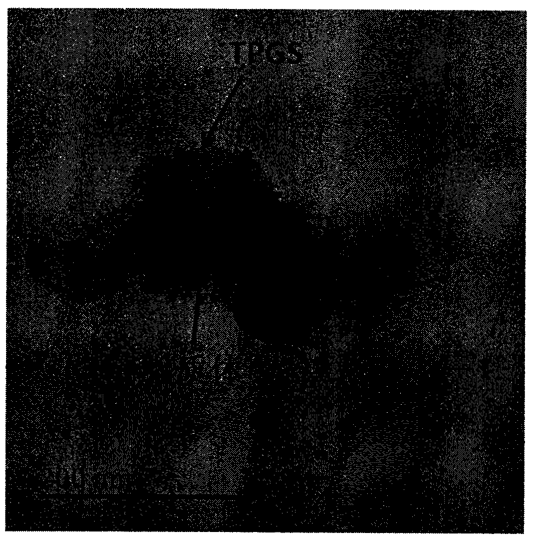
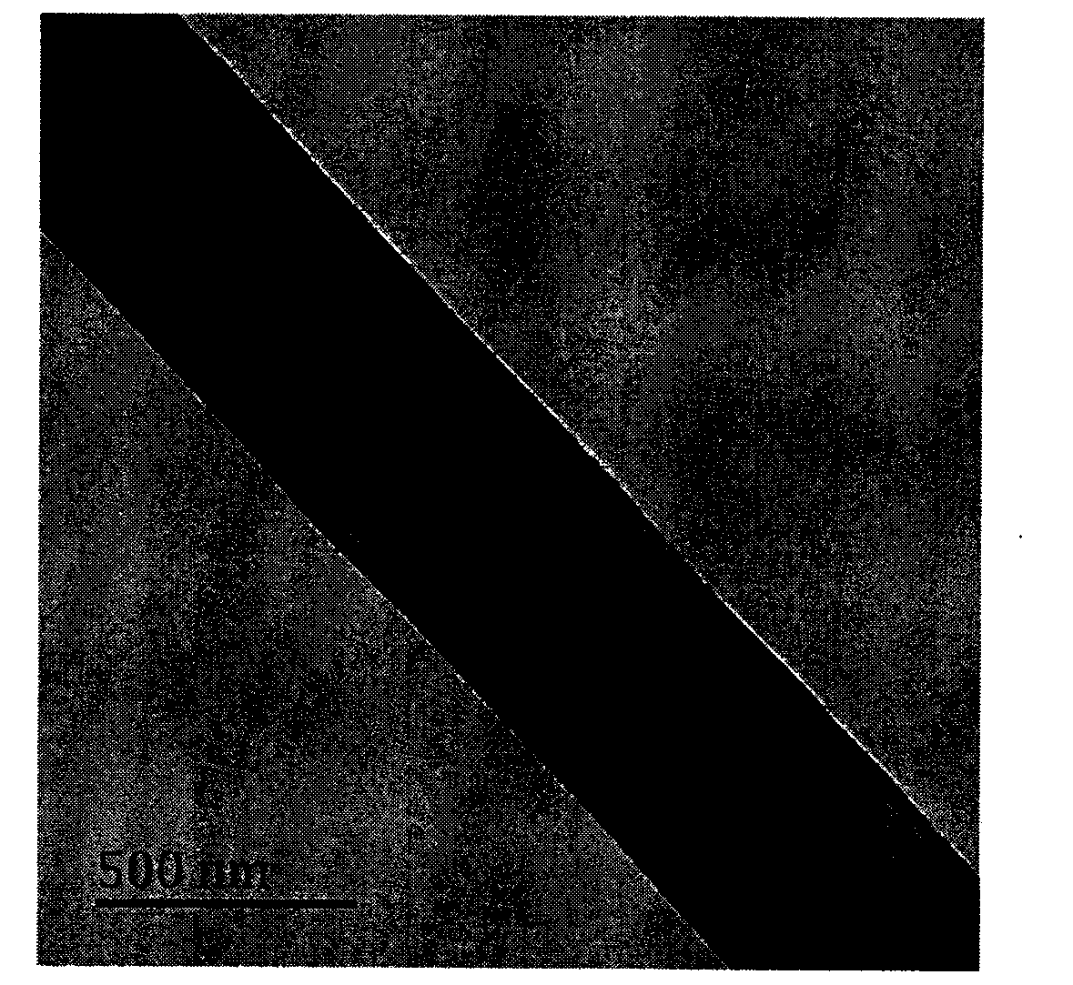
[0032] Step 1: film dispersion method to prepare drug-loaded liposomes modified by TPGS: take an appropriate amount of lecithin, cholesterol and doxorubicin (mass ratio 20:2:1) and add them to a round bottom flask, then add absolute ethanol, After stirring and dissolving at room temperature, place it on a rotary evaporator to completely remove ethanol and obtain a lipid film; prepare a 0.5% (mass volume ratio) TPGS aqueous solution, add it to the above lipid film, stir at room temperature for 2 hours, and use Ultrasonic cell pulverizer ultrasonically obtains the drug-loaded liposome suspension modified by TPGS with uniform particle size; the TEM picture of the prepared drug-loaded liposome modified by TPGPS is as follows: figure 1 shown. TPGS is wrapped around drug-loaded liposomes in cloudy form, and the multilayer structure o...
Embodiment 2
[0036] The preparation of the TPGS liposome-fish gelatin composite nanofiber scaffold loaded with epidermal growth factor consists of the following three steps:
[0037] Step 1: Preparation of drug-loaded liposomes modified by TPGS by film dispersion method: Weigh lecithin, cholesterol and epidermal growth factor (mass ratio 20:2:1) into a round bottom flask, then add absolute ethanol, room temperature After stirring and dissolving, place it on a rotary evaporator to completely remove ethanol and obtain a lipid film; prepare a 0.5% (mass volume ratio) TPGS aqueous solution, add it to the above lipid film, stir at room temperature for 2 hours, and use ultrasonic The cell pulverizer ultrasonically obtains the drug-loaded liposome suspension modified by TPGS with uniform particle size;
[0038] Step 2: Prepare an aqueous solution containing 20% (mass volume ratio) fish gelatin, and add the drug-loaded liposome suspension modified by TPGS prepared in step 1 in a ratio of 10%-20%...
PUM
 Abstract
Description
Claims
Application Information
 - Generate Ideas
- Intellectual Property
- Life Sciences
- Materials
- Tech Scout
- Unparalleled Data Quality
- Higher Quality Content
- 60% Fewer Hallucinations
Browse by: Latest US Patents, China's latest patents, Technical Efficacy Thesaurus, Application Domain, Technology Topic, Popular Technical Reports.
© 2025 PatSnap. All rights reserved.Legal|Privacy policy|Modern Slavery Act Transparency Statement|Sitemap|About US| Contact US: help@patsnap.com