Reactive-oxygen-species sensitive nanoparticle capable of promoting vascularization of surface of wound and preparation method thereof

A technology of nanoparticles and reactive oxygen species, which is applied in the field of medicine to maintain protein activity and promote vascularization of wounds

Inactive Publication Date: 2015-05-06
SECOND MILITARY MEDICAL UNIV OF THE PEOPLES LIBERATION ARMY
View PDF2 Cites 9 Cited by
  • Summary
  • Abstract
  • Description
  • Claims
  • Application Information

AI Technical Summary

Problems solved by technology

[0006] At present, there is no literature reporting a nanoparticle that is sensitive to reactive oxygen species, ca

Method used


Image

  • Reactive-oxygen-species sensitive nanoparticle capable of promoting vascularization of surface of wound and preparation method thereof
  • Reactive-oxygen-species sensitive nanoparticle capable of promoting vascularization of surface of wound and preparation method thereof
  • Reactive-oxygen-species sensitive nanoparticle capable of promoting vascularization of surface of wound and preparation method thereof

Examples


Embodiment 1

[0041] Embodiment 1: Preparation of drug-loaded nanoparticles of the present invention

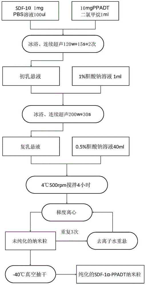
[0042] Such as image 3 As shown, the w1 / o / w2 method is applied, 1mg murine SDF-1α is dissolved in 100ulPBS solution as the inner water phase, 10mgPPADT is dissolved in 1ml dichloromethane as the oil phase, and the initial milk suspension. The colostrum suspension was added to 1ml of 1% sodium cholate solution (outer aqueous phase), and the double emulsion suspension was formed by continuous ultrasonication at 200 watts*30s. Then put the double emulsion suspension into 40ml of 0.5% sodium cholate solution and stir at 500rpm at 4°C for 4 hours to remove the organic solvent. Nanoparticles were collected after gradient centrifugation, resuspended in deionized water, and repeated three times to remove SDF-1α and sodium cholate in the solution to obtain SDF-1α-PPADT nanoparticles.

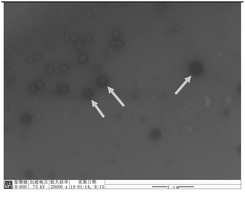
[0043] The number of nanoparticles measured by the particle size analyzer and the transmission electron micros...

Embodiment 2

[0044] Embodiment 2: SDF-1α-PPADT release experiment in vitro

[0045] Dissolve the nanoparticles prepared in the previous step with 1ml of dichloromethane, centrifuge at 12000g for 10 minutes, remove the supernatant, dissolve the SDF-1α attached to the wall with an appropriate amount of PBS solution, and measure its content by ELISA. When the dosage of SDF-1α is 1 mg and the dosage of PPADT is 10 mg, each 1 mg of nanoparticles contains 18ug of SDF-1α. 100 mg of SDF-1α-PPADT nanoparticles were resuspended in 1 ml of deionized water and Fenton’s reagent (3% H 2 o 2 Add 1% ferrous chloride), take an appropriate amount of suspension respectively at 30min, 1h, 3h, 6h, 12h, 24h, 48h, remove nanoparticles by centrifugation at 20000g for 10min, and measure the concentration of SDF-1α in the supernatant by ELISA. In aqueous solution, nanoparticles released 5% of the total amount of SDF-1α encapsulated within 48h. In the presence of oxygen free radicals, SDF-1α in the nanoparticles ...

Embodiment 3

[0047] Embodiment 3: PPADT cytotoxicity experiment

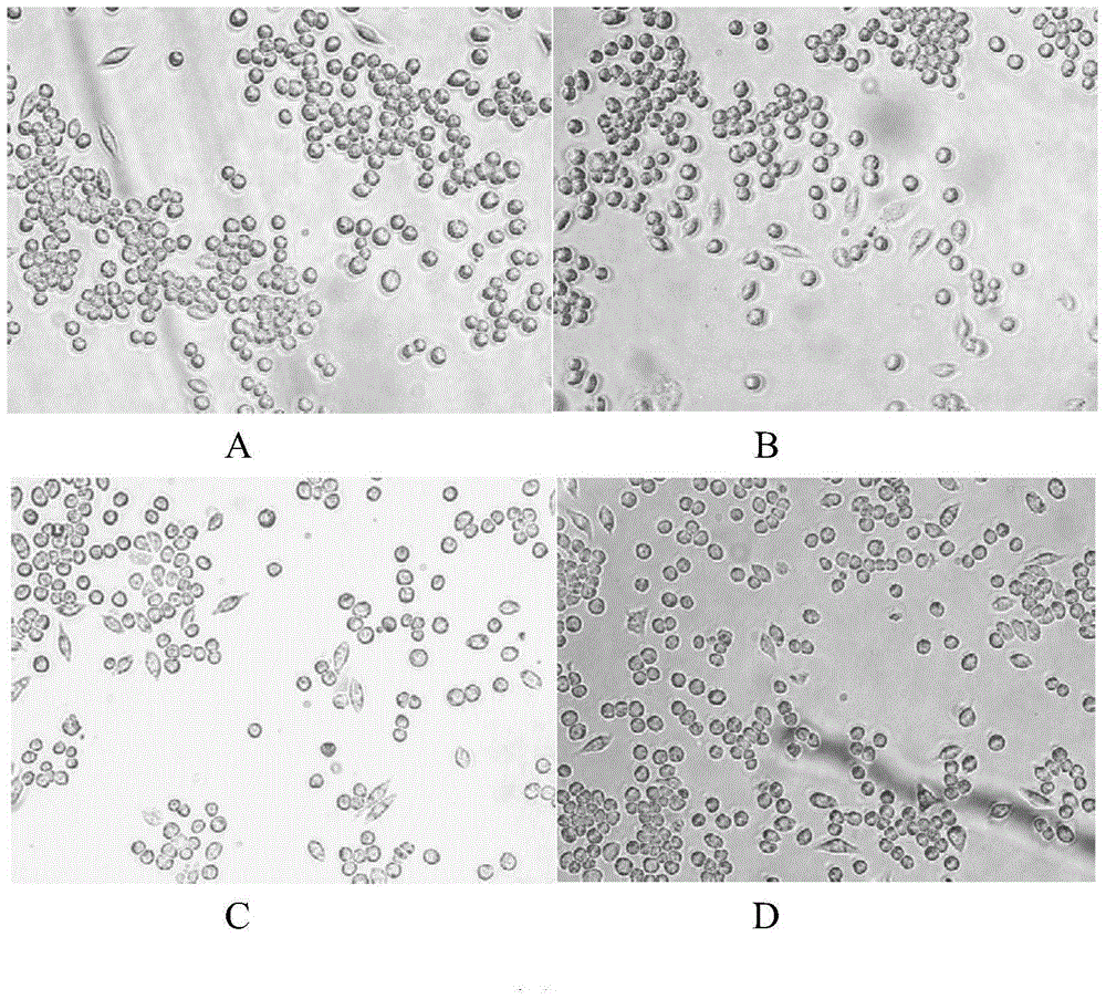
[0048] The low-glucose DMEM cell culture with 10% fetal bovine serum is based on culturing RAW264.7 in a 6-well plate, and the cell density is 1X10 5 / well, add deionized water, 1mg / ml blank nanoparticles, 10mg / ml blank nanoparticles, 100mg / ml blank nanoparticles to culture for 24 hours, 6 wells in each group, stain with CCK8 after 24 hours, under 200X microscope Observe 10 fields of view, count the number of surviving cells, and count the absorbance value at 570nm, and compare the cell proliferation rates of each group. It is found that after 24 hours of co-culture with PPADT nanoparticles and RAW264.7 cells, the cell proliferation rate is basically the same as that of the control group. similar (Table 1, figure 2 ). Prove that the cytotoxicity of nanoparticles is weak.

[0049] Table 1: Cell Viability Detection

[0050] (cytotoxicity test co-cultured for 24 hours CCK8 staining, relative absorbance value)

[0051] ...


PUM

PropertyMeasurementUnit
Particle sizeaaaaaaaaaa

Abstract

The invention belongs to the technical field of medicines and discloses a reactive-oxygen-species sensitive nanoparticle capable of promoting the vascularization of the surface of a wound and a preparation method thereof. The active component of the nanoparticle is a stromal cell derived factor 1 alpha (SDF-1alpha), and the medicine carrying nanomaterial is poly(1,4-phenyleneacetone dimethylenethioketal) (PPADT). The nanoparticle has a size of 70-130nm. The invention further discloses the preparation method of the nanoparticle. The medicine carrying nanoparticle is prepared by adopting a multiple emulsion-solvent evaporation method. The product has a good slow-releasing effect, is reduced in protein denaturation and improved in encapsulation rate and has good stability and low biotoxicity.

Description

technical field [0001] The invention relates to the technical field of medicine, in particular to a nanoparticle sensitive to active oxygen and capable of promoting wound surface vascularization and a preparation method thereof. Background technique [0002] Wound repair is a complex biological system engineering. In the process, a variety of cells, growth factors and cytokines participate together. Through changes in a variety of cellular and molecular levels, the barrier function of the skin is reconstructed (1. Balaji, Sundeep G. Keswani , and Timothy M. Crombleholme. The Role of Mesenchymal Stem Cells in the Regenerative Wound Healing Phenotype, ADVANCES IN WOUND CARE, VOLUME 1, NUMBER 4, 2012:159-165). Among them, bone marrow mesenchymal stem cells (BMSCs), as a kind of cells with multipotential differentiation potential (2. Singer NG and Caplan AI. Mesenchymal stem cells: mechanisms of inflammation. Annu Rev Pathol 2011; 6:457.), can participate in Vascularization and...

Claims


Application Information

Patent Timeline
IPC IPC(8): A61K38/19A61K47/34A61K9/14A61P17/02A61L15/44A61L15/26A61L15/32
Inventor 王光毅姜浩汤焘纪世召何放夏照帆
Owner SECOND MILITARY MEDICAL UNIV OF THE PEOPLES LIBERATION ARMY
Why Eureka
Try Eureka