Fungus fluorescence staining method and application thereof

A fluorescent dyeing and fungal technology, which is applied in the field of fluorescent dyeing, can solve the problems of time-consuming, high technical requirements for operators, and inability to express green fluorescent protein genes, etc., and achieve the effect of simple operation method and simplified dyeing steps

Inactive Publication Date: 2015-09-09
HUNAN AGRICULTURAL UNIV
View PDF2 Cites 4 Cited by
  • Summary
  • Abstract
  • Description
  • Claims
  • Application Information

AI Technical Summary

Problems solved by technology

However, this method involves the transgenic operation process, including the construction of the expression vector and the genetic transformation process, which takes longer and requires higher technical r

Method used


Image

  • Fungus fluorescence staining method and application thereof
  • Fungus fluorescence staining method and application thereof
  • Fungus fluorescence staining method and application thereof

Examples


Embodiment 1

[0028] Take the invasion process of potato late blight as an example.

[0029] 1. Materials

[0030] Potato variety and physiological race of Phytophthora infestans: The tested potato variety was Fevorita, and Phytophthora infestans was from Hunan Agricultural University.

[0031] 2. Method

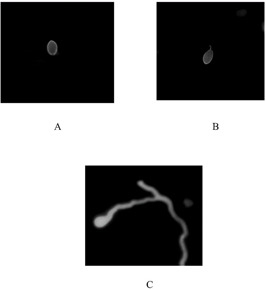
[0032] Sow the tubers of the tested potato varieties in small flowerpots, and grow seedlings under the conditions of 16-20°C and 12-16 hours of sunshine. After flattening the leaves of the seedlings, use a concentration of about 1×10 5 Spray inoculation with sporangia suspension of P. infestans infestans per mL, and then move the flower pot into an artificial climate box for cultivation. The cultivation conditions are: 16-18°C, 16 hours of light, light intensity not less than 10000lx, and humidity of 100%.

[0033] The leaves were sampled at 0h, 12h, 24h, 48h, 72h, and 96h after inoculation.

[0034] The leaves of 0h, 12h, and 24h after inoculation were directly dyed, that is, the dye...

Embodiment 2

[0037] Coleomyces cowpea ( Cercospora vignae ) invasion of cowpea as an example.

[0038] The cowpea coal mold fungus came from the laboratory of the Plant Protection College of Hunan Agricultural University. The variety of cowpea is red-billed swallow, which was purchased from the Institute of Vegetables and Flowers, Hunan Academy of Agricultural Sciences.

[0039] The cowpea leaves were sampled at about 0h-5h and 72h after inoculation with cowpea coal mold fungus.

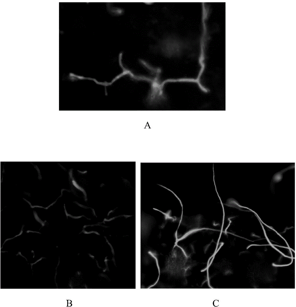
[0040] The leaves of 0-5h after inoculation are directly dyed, that is, the dyeing solution is evenly sprayed on the surface of the leaves, and then the UV-2A filter is configured (excitation filter, 330-380nm; dichroic mirror, 400nm; barrier filter 420nm) ) under a fluorescence microscope (do not add a cover glass) to observe the situation of cowpea coal mold spores on the surface of cowpea leaves, see image 3 a.

[0041] About 72 hours after inoculation, the leaf material is first decolorized and then dye...

Embodiment 3

[0043] Eggplant anthracnose bacteria ( Colletotrichum truncatum ) invasion of eggplant as an example.

[0044] Eggplant anthracnose bacteria came from the Laboratory of Plant Protection College of Hunan Agricultural University. The eggplant variety was Heiguan Zaoqie, which was purchased from the Institute of Vegetables and Flowers, Hunan Academy of Agricultural Sciences.

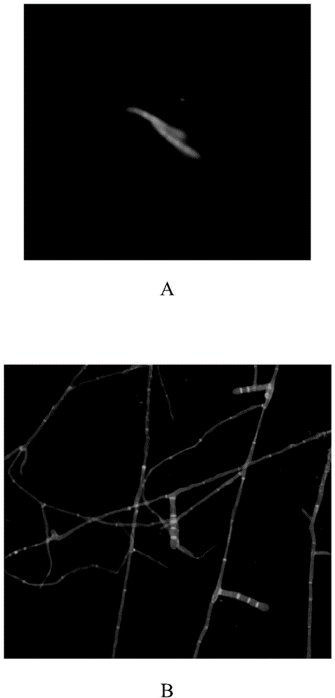
[0045] The eggplant leaves were sampled at about 0h-5h and 72h after inoculation with Eggplant anthracnose bacteria.

[0046] The staining method is the same as that in Example 2, and the eggplant anthracnose spores are observed under a fluorescence microscope on the eggplant leaf surface ( Figure 4 A) and mycelia of P. anthracnose in eggplant leaf tissue ( Figure 4 B) Situation.


PUM


Abstract

The invention discloses a fungus fluorescence staining method. The method includes firstly, spraying a staining solution [dissolving direct yellow 96 in a 0.1M Tris-Hcl buffer solution with the pH (potential of hydrogen) of 8.5] on the surfaces of host plant leaves uniformly directly to observe conditions of pathogenic fungal spores on the surfaces of the host plant leaves; secondly, soaking leaves decayed by pathogenic fungal hyphae into 95% (v/v) ethyl alcohol in boiling water bath to remove chlorophyll completely; thirdly, taking out the leaves, and cleaning the leaves by 50% (v/v) ethyl alcohol, 50mM sodium hydroxide and pure water sequentially; finally, soaking the leaves into the staining solution to stain so as to observe the conditions after pathogenic fungi invade host leaf tissues. The fungus fluorescence staining method has the advantages that all reagents used in the method are poisonless and harmless and are safer and more environment friendly than reagents such as chloroform, phenol, trichloroacetic acid and pyridine used in conventional decoloration methods; the fungus fluorescence staining method is simple and feasible, and a slicing process is omitted and staining steps are simplified on the premise of not affecting the staining effect.

Description

technical field [0001] The invention relates to a fluorescent dyeing method, in particular to a fungal fluorescent dyeing method, and also relates to the application of the method in observing the invasion process of pathogenic fungi. Background technique [0002] In the study of phytopathology, it is often necessary to observe the interaction between pathogenic fungi and host plants. The traditional tissue transparent staining method can quantitatively study the expansion of pathogen hyphae in tissues and reveal the spatiotemporal dynamics of host-pathogen interactions. However, the application of this tissue transparent staining method has certain limitations: first, it is difficult to distinguish between pathological necrosis and mechanical trauma of cells, which will lead to unsatisfactory observation results; second, it is not suitable for observing the germination and invasion of fungal spores on the surface of host plants. The process, the tissue transparent staining...

Claims


Application Information

Patent Timeline
IPC IPC(8): C12Q1/02G01N1/30
Inventor 周倩刘甜甜熊兴耀
Owner HUNAN AGRICULTURAL UNIV
Why Eureka
Try Eureka