Nanometer vesica capable of detecting wild type p53 protein and variant p53 protein in cells simultaneously

A nanovesicle and wild-type technology, applied in measurement devices, material analysis through optical means, instruments, etc., can solve problems such as low sensitivity, large staining differences, and inability to analyze and detect p53 protein in situ, achieving Improved specificity and sensitivity, high specificity effect

Inactive Publication Date: 2015-10-14
EAST CHINA UNIV OF SCI & TECH
View PDF12 Cites 10 Cited by
  • Summary
  • Abstract
  • Description
  • Claims
  • Application Information

AI Technical Summary

Problems solved by technology

Immunohistochemistry is widely used, but its main disadvantage is that the staining varies greatly among different batches and the repeatability is poor.
The ELISA method uses antigen-antibody sandwich reaction to detect the target protein, which has good stability, but the method is cumbersome and the sensitivity is not high
Th

Method used


Image

  • Nanometer vesica capable of detecting wild type p53 protein and variant p53 protein in cells simultaneously
  • Nanometer vesica capable of detecting wild type p53 protein and variant p53 protein in cells simultaneously

Examples


Embodiment 1

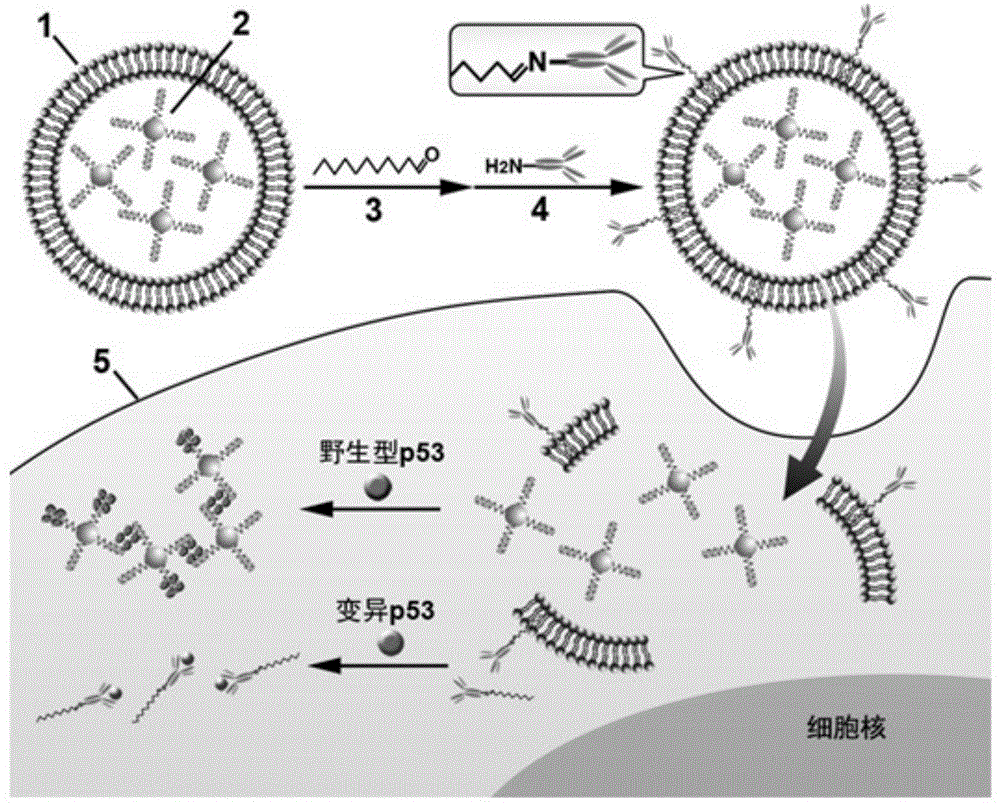
[0019] Example 1. Synthesis of nanovesicles that can simultaneously detect wild-type and mutant p53 proteins in cells

[0020] (1) According to the existing literature, 13nm gold colloid was synthesized as a seed. 50mL HAuCl 4 (0.01%) solution was added to the round bottom flask and heated to boiling, then 5mL sodium citrate solution (38.8mM) was added rapidly and continued to stir for 30 minutes until the solution turned red, and the obtained 13nm gold seeds were used to prepare the particle size in Nano gold around 60nm.

[0021] (2) Using gold species to synthesize 60nm gold nanoparticles. Mix 1 mL of gold seed solution with 100 µL of NH 2 OH·HCl (0.2M) was mixed and diluted to 25mL with pure water, then 3.0mLHAuCl 4 (0.01%) was slowly added dropwise into the above mixed solution, and the stirring was continued for 30 minutes until the solution turned dark red. The obtained nano-gold solution was centrifuged, washed with pure water, and stored at 4°C.

[0022] (3) Usi...

Embodiment 2

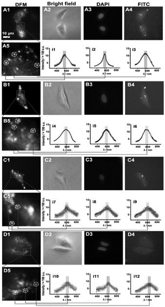
[0024] Example 2. In situ imaging and detection of the distribution of p53 protein in cells using nanovesicles

[0025] Using the HeLa cervical cancer cell line as a model, first HeLa cells (1.0 mL, 1×10 6 mL -1 ) were planted in confocal culture dishes and cultured for 24 hours. Then 60 μL of nanovesicles were added to the dish and incubated at 37°C for 2 hours. The dishes were washed with PBS and the cells were submerged, then observed under a dark-field microscope and a fluorescence microscope. Cell imaging see figure 2 . It was found that in the tumor cell HeLa, the color of gold nanoparticles in the dark field image was green, and there was no obvious aggregation, indicating that the content of wild-type p53 was low; while the fluorescence signal of FITC was strong, indicating that the content of mutant p53 was high. With nutlin-3 drug solution (250μg mL -1 ) to cells (1.0mL, 1×10 6 mL -1 ) for pretreatment, cells were co-cultured with drugs for 48 hours, then 60...


PUM

PropertyMeasurementUnit
Particle sizeaaaaaaaaaa

Abstract

The invention discloses a nanometer vesica capable of detecting wild type p53 protein and variant p53 protein in cells simultaneously. Nanogold capable of specifically recognizing wild type p53 protein wraps inside the vesica, and an antibody resisting variant p53 protein marked by fluorescein isothiocyanate decorates the surface of the vesica. The nanometer vesica has the advantages that (1) the nanometer vesica recognizes different types of tumor suppressor proteins p53 with high specificity, and the detection specificity and the sensitivity are improved; (2) plasma resonance imaging and fluorescence imaging technologies are adopted, after the vesica enters the cell, the cell is subjected to in-situ imaging by adopting a microscope, and wild type p53 protein and variant p53 protein on the single-cell layers are detected simultaneously; (3) the nanometer vesica designed according to the technical scheme is capable of carrying out in-situ monitoring on variation of expression of p53 protein in the cell under the action of medicine, and a novel method is provided for researching physiological process in the cell participated by the vesica.

Description

technical field [0001] The invention relates to the fields of cell live imaging analysis technology, dark field microscopy technology and nano material preparation, in particular to a nano vesicle capable of simultaneously detecting intracellular wild-type and variant p53 proteins and its application. Background technique [0002] In recent years, plasmonic nanoparticles have attracted extensive attention. These nanoparticles have special optical and physical properties, can effectively absorb and scatter visible light, and induce metal surface plasmon resonance. Thanks to the development of dark-field microscopy and plasmon resonance Rayleigh scattering spectroscopy, plasmonic nanoparticles have been successfully used in many research fields such as biosensing, single particle analysis, and drug delivery. Studies have shown that plasmonic nanoprobes have become a powerful tool for studying living organisms, especially cells, and can be used for cell imaging, cell surface re...

Claims


Application Information

Patent Timeline
IPC IPC(8): G01N21/552G01N21/64
Inventor 龙亿涛钱若灿曹玥
Owner EAST CHINA UNIV OF SCI & TECH
Why Eureka
Try Eureka