Preparation method and application of metal sulfur-group compound multifunctional nano probe

A nano-probe and compound technology, applied in the fields of material chemistry and biomedicine, can solve the problems of ultra-small cuprous selenide nanoparticles that have not been reported, and achieve broad market application prospects and mild preparation methods

Active Publication Date: 2016-07-13
SUZHOU UNIV
View PDF4 Cites 6 Cited by
  • Summary
  • Abstract
  • Description
  • Claims
  • Application Information

AI Technical Summary

Problems solved by technology

However, ultrasmall cuprous selenide nanoparticles (less than 10 nm) with good biocompatibility and water solubility for multimodality-mediated photothermal therapy have not yet been reported.

Method used


Image

  • Preparation method and application of metal sulfur-group compound multifunctional nano probe
  • Preparation method and application of metal sulfur-group compound multifunctional nano probe
  • Preparation method and application of metal sulfur-group compound multifunctional nano probe

Examples


Embodiment 1

[0046] Disperse 0.5 mmol of selenium powder in 50 mL of deionized water, add 1.5 mmol of sodium borohydride powder into the selenium powder dispersion system under the protection of an inert gas, mix well, and reduce the selenium powder to colorless. 1 g of mercaptosuccinic acid and 0.17 g of copper chloride dihydrate powder were dissolved in 5 mL of water, and then added to the above-mentioned selenium precursor solution, and stirred and reacted at room temperature for 1 hour. After the reaction is finished, the solution is subjected to multiple ultrafiltration separations to obtain cuprous selenide nanoparticles stabilized by mercaptosuccinic acid.

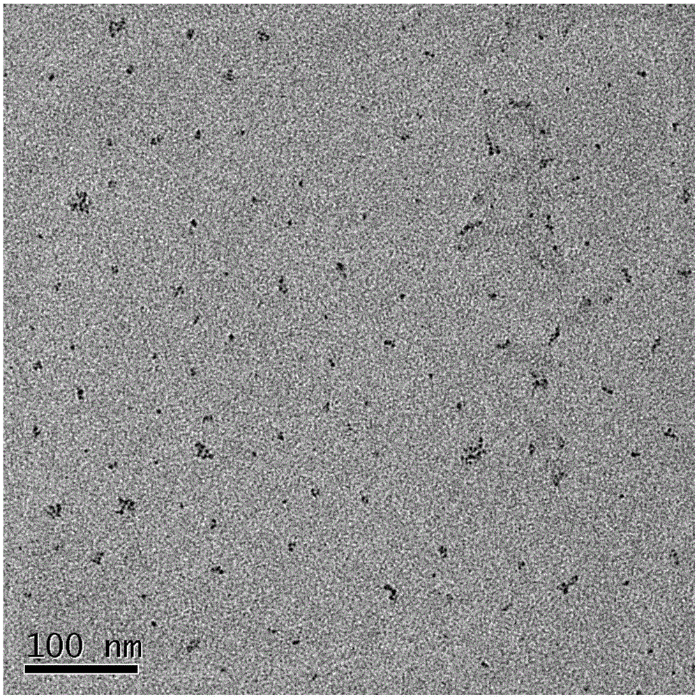
[0047] The cuprous selenide sample stabilized by mercaptosuccinic acid was redispersed in about 30mL of aqueous solution, and 0.2g of bismercaptopolyethylene glycol (molecular weight: 5000) was added. After stirring and reacting at room temperature for 6 hours, the solution was separated by ultrafiltration several times. After d...

Embodiment 2

[0053] Disperse 0.5mmol of selenium powder in 50mL of deionized water, and pass through inert gas protection. Add 1.5 mmol of sodium borohydride powder into the selenium powder dispersion system, mix well, and reduce the selenium powder to colorless. 1 g of polyvinylpyrrolidone (PVP-K30) and 0.17 g of copper chloride dihydrate powder were dissolved in 5 mL of water, and then added to the above-mentioned selenium precursor solution, and stirred for 1 hour at room temperature. After the reaction is finished, the solution is subjected to multiple ultrafiltration separations to obtain a polyvinylpyrrolidone-stabilized cuprous selenide sample.

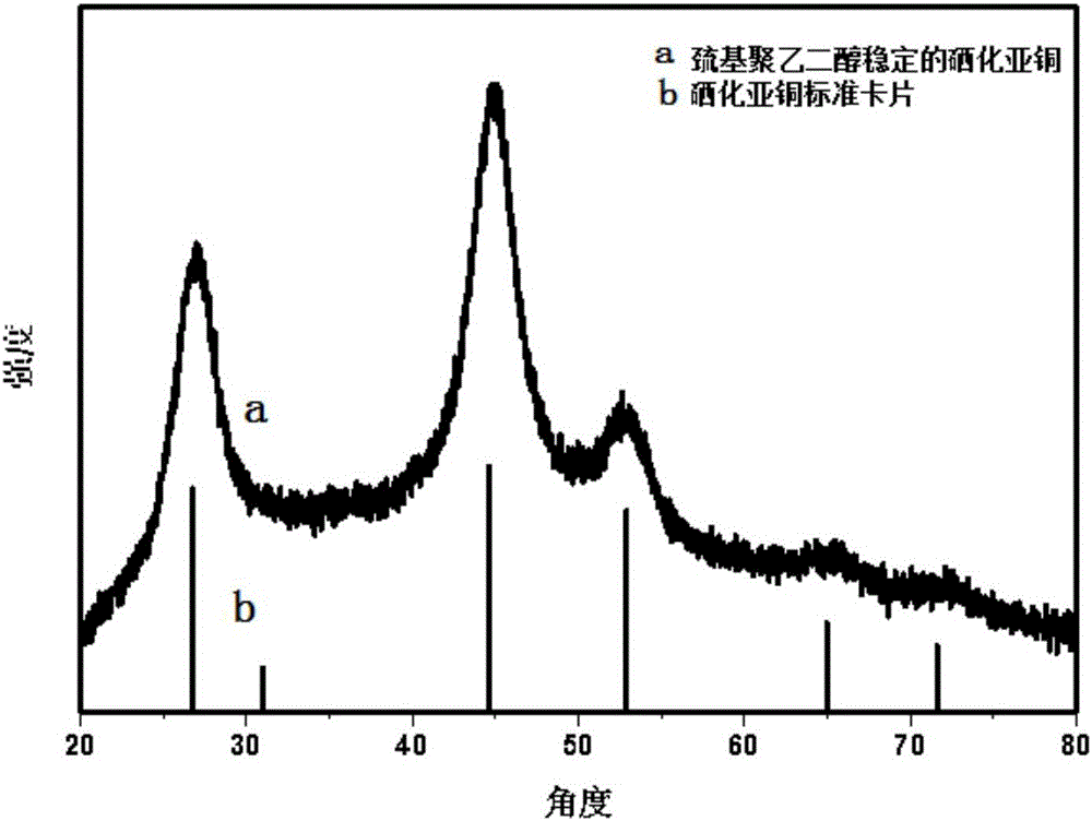
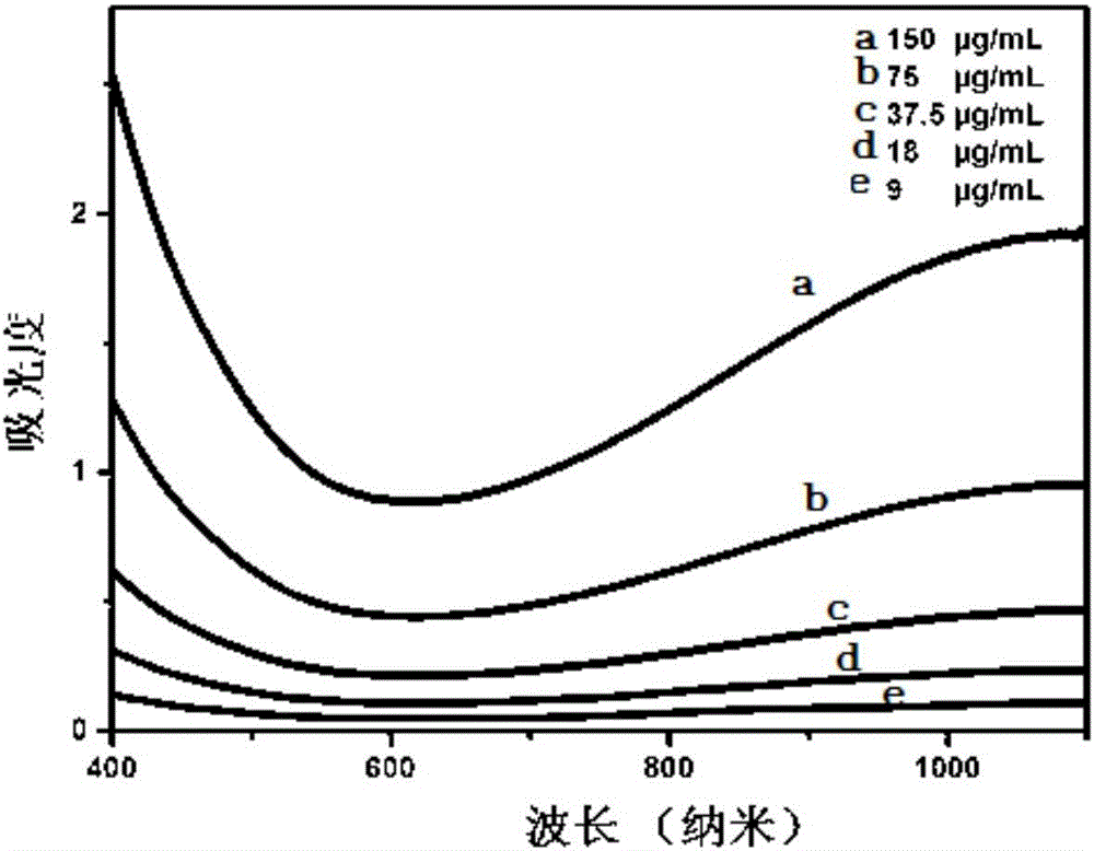
[0054] attached Figure 7 X-ray diffraction pattern of cuprous selenide nanoparticles stabilized by polyvinylpyrrolidone. attached Figure 8 It is the UV absorption pattern of cuprous selenide nanoparticles stabilized by water-soluble polyvinylpyrrolidone. attached Figure 9 It is a transmission electron micrograph of cuprous selenide ...

Embodiment 3

[0056] Disperse 0.5mmol of selenium powder in 50mL of deionized water, and pass through inert gas protection. Add 1.5 mmol of sodium borohydride powder into the selenium powder dispersion system, mix well, and reduce the selenium powder to colorless. 1 g of mercaptosuccinic acid and 0.17 g of silver nitrate powder were dissolved in 5 mL of water, then added to the above-mentioned selenium precursor solution, and stirred and reacted at room temperature for 1 hour. After the reaction is finished, the solution is separated by ultrafiltration several times to obtain a silver selenide sample stabilized by mercaptosuccinic acid.

[0057] attached Figure 10 X-ray diffraction pattern of silver selenide nanoparticles stabilized with mercaptosuccinic acid. attached Figure 11 UV absorption pattern of silver selenide nanoparticles stabilized by water-soluble mercaptosuccinic acid. attached Figure 12 It is a transmission electron micrograph of silver selenide nanoparticles.


PUM

PropertyMeasurementUnit
sizeaaaaaaaaaa

Abstract

The invention belongs to the field of material chemistry and biological medicine, and in particular relates to a preparation method and application of a metal sulfur-group compound multifunctional nano probe. The preparation method comprises the following steps: in the presence of a water-soluble sulfhydryl compound or a biocompatible molecule, enabling low-valence sulfur group anions to have stirred mixing reaction with metal cations in an aqueous solution; and then, centrifuging, carrying out ultrafiltration, modifying, dialyzing and purifying to obtain water-soluble metal sulfur-group compound nanoparticles which can be used for photoacoustic imaging, CT imaging, nuclear medical imaging and photothermal therapy. The preparation method is gentle, cheap, quick and efficient; and the obtained nano microparticles are small in size, can circulate for a relatively long time in vivo, and can be gathered in tumors by enhancing permeation and retention effects.

Description

technical field [0001] The invention belongs to the fields of material chemistry and biomedicine, and in particular relates to a preparation method and application of a metal chalcogenide multifunctional nanometer probe. Background technique [0002] Cancer is the leading cause of human death worldwide. Photothermal therapy, as a minimally invasive, efficient and promising cancer treatment method, has attracted extensive attention from researchers. Photothermal therapy is the use of photothermal therapeutic agents to absorb near-infrared light to generate thermal effects to kill tumor cells. During the treatment process, it is necessary to combine medical imaging technology to monitor the therapeutic effect. Therefore, through the research and development, optimization and upgrading of nanomaterials for photothermal therapy, the integration of contrast agents with imaging enhancement effects and photothermal therapeutic agents is of great significance for the realization of...

Claims


Application Information

Patent Timeline
Patent Type & Authority Applications(China)
IPC IPC(8): C09K11/02C09K11/88B82Y30/00B82Y40/00A61K41/00A61K51/06
CPCA61K41/0052A61K51/06B82Y30/00B82Y40/00C09K11/025C09K11/881
Inventor 李桢张少华
Owner SUZHOU UNIV
Why Eureka
Try Eureka