
A preparation method and application of decellularized amniotic membrane for skin refractory wound repair

A wound repair and decellularization technology, applied in animal cells, vertebrate cells, artificial cell constructs, etc., can solve the problems of amniotic matrix damage, basement membrane component damage, basement membrane damage, etc., to promote regeneration and repair, ensure Integrity, effect of improving decellularization efficiency

Active Publication Date: 2019-05-10
天晴干细胞股份有限公司
View PDF5 Cites 0 Cited by
  • Summary
  • Abstract
  • Description
  • Claims
  • Application Information

AI Technical Summary

Problems solved by technology

However, the existing amniotic membrane decellularization method still cannot guarantee the integrity of the amniotic matrix on the basis of complete removal of cells, especially it will cause serious damage to the basement membrane, affecting the adhesion and migration of seed cells or endogenous cells in vivo
Patent Publication No. CN1757717A (preparation method of dry active amniotic membrane) uses trypsin to treat for a long time in combination with cell scraper or cotton swab to remove epithelial cells, but long-term trypsin digestion will lead to changes or even degradation of amnion collagen structure, while physical scraping Injury to basement membrane components that severely disrupt amnion epithelial cell adhesion
Patent Publication No. CN102327640A (a preparation method of decellularized amnion) uses SDS for cell lysis to promote the detachment of amnion epithelial cells, but low concentration of SDS will limit the removal efficiency of amnion cells, but high concentration of SDS will cause damage to the amnion matrix

Method used

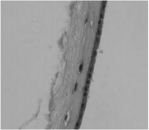
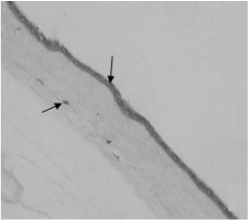
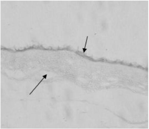

Image

  • A preparation method and application of decellularized amniotic membrane for skin refractory wound repair
  • A preparation method and application of decellularized amniotic membrane for skin refractory wound repair
  • A preparation method and application of decellularized amniotic membrane for skin refractory wound repair

Examples


specific Embodiment approach 1

[0033] Specific embodiment 1: A method for preparing decellularized amniotic membrane for repairing difficult-to-heal skin wounds according to this embodiment is characterized in that it is performed according to the following steps:

[0034] 1. Choose the full-term placenta, bluntly strip the amniotic membrane tissue, wash it with normal saline 2 to 3 times to remove blood clots, colloids and other impurities, and then soak it in normal saline containing antibiotics for 10 minutes for disinfection;

[0035] 2. Lay the amniotic membrane with the epithelium side up on the sterile nitrocellulose membrane, and let it dry for 1 to 2 hours under aseptic conditions to make the amniotic membrane and the nitrocellulose membrane closely adhere to form an amniotic membrane patch and cut it to an appropriate size size;

[0036] 3. Place the amniotic membrane patch in the mixed digestion solution with a concentration of 0.25%~0.5% trypsin and 0.2~0.5g / LEDTA·4Na by mass volume percentage, and sha...

specific Embodiment approach 2

[0044] Specific embodiment two: This embodiment is different from specific embodiment one in that the antibiotic content in the antibiotic-containing physiological saline described in step one is: penicillin with a concentration of 50 mg / L and streptomycin with a concentration of 50 mg / L And amphotericin B at a concentration of 2.5 mg / L. Others are the same as the first embodiment.

specific Embodiment approach 3

[0045] Specific embodiment three: This embodiment is different from specific embodiment one in that the open time in step two is 1 to 1.5 hours. Others are the same as the first embodiment.


PUM


Abstract

The invention relates to a preparing method and application of an amniotic membrane, in particular to a preparing method and application of an acellular amniotic membrane used for repairing skin wound difficult to heal. The preparing method specifically comprises the steps of isolating, cleaning and sterilizing an amniotic membrane tissue; arranging and drying the amniotic membrane tissue on a nitrocellulose membrane to manufacture an amniotic membrane paster; vibrating and digesting the amniotic membrane paster in mixed digestive fluid of 0.25%-0.5% of pancreatin and 0.2-0.5g / L EDTA.4Na for 10-30 minutes at a temperature of 37 DEG C; rinsing the amniotic membrane paster in TE buffer solution; vibrating overnight to fully remove amniotic membrane cells in TE-TritonX-100 solution; washing the amniotic membrane paster for three times in TE solution; vibrating and degrading the amniotic membrane paster for 2-4 hours in nucleic acid degrading solution at a temperature of 37 DEG C; washing the amniotic membrane paster for three times in the TE solution. The prepared acellular amniotic membrane paster can be stored for a long time after being refrigerated, dried, packed and sterilized by cobalt 60 irradiation and is a tissue engineering material which can be taken and used when needed. The acellular amniotic membrane paster can be easily separated from the nitrocellulose membrane to be used for repairing the skin wound after rewatered.

Description

Technical field [0001] The invention relates to a preparation method and application of amniotic membrane. Background technique [0002] Amniotic membrane tissue discarded during the perinatal period is one of the important sources of seed cells for regenerative medicine. It has a wide range of sources, is easy to obtain, and has no ethical and moral controversy. Amniotic membrane, as an immune-exempt biological graft, has biological properties such as anti-inflammatory, anti-adhesion, pain relief, and promotion of epithelialization. It has been used in burn patients and other surgical procedures since 1910 to promote epidermalization, Relieve pain and prevent infection. In recent years, with the further understanding of amniotic membrane and the development of amniotic membrane preservation and preparation technology, amniotic membrane has once again become a focus of research and application, and is widely used in corneal reconstruction, plastic surgery, burn treatment and oth...

Claims


Application Information

Patent Timeline
Patent Type & Authority Patents(China)
IPC IPC(8): C12N5/071A61L27/36A61L27/60
CPCA61L27/3604A61L27/3687A61L27/3691A61L27/60A61L2430/40
Inventor 付寅生张怡孟庆雪刘艳青
Owner 天晴干细胞股份有限公司
Learn More