Placental villus plate mesenchymal stem cells and clinical preparation method

A technique for stromal stem cells and villous plate, which is applied in the field of placental villus plate mesenchymal stem cells and clinical preparation, can solve the problems of lowering production cost and need to be further improved, and achieves easier control of process stability, reduction of cell consumption, and avoidance of dryness. Sex-reducing effect

Inactive Publication Date: 2016-11-16
四川华皓生物科技有限公司
View PDF3 Cites 4 Cited by
  • Summary
  • Abstract
  • Description
  • Claims
  • Application Information

AI Technical Summary

Problems solved by technology

Chinese patent CN102586184A discloses a method for establishing a placental mesenchymal dry cell bank, which uses tissue digestion enzyme solution (containing dispase, trypsin, deoxyribonuclease I, collagenase IV, hyaluro...

Method used


Image

  • Placental villus plate mesenchymal stem cells and clinical preparation method
  • Placental villus plate mesenchymal stem cells and clinical preparation method
  • Placental villus plate mesenchymal stem cells and clinical preparation method

Examples


Embodiment 1

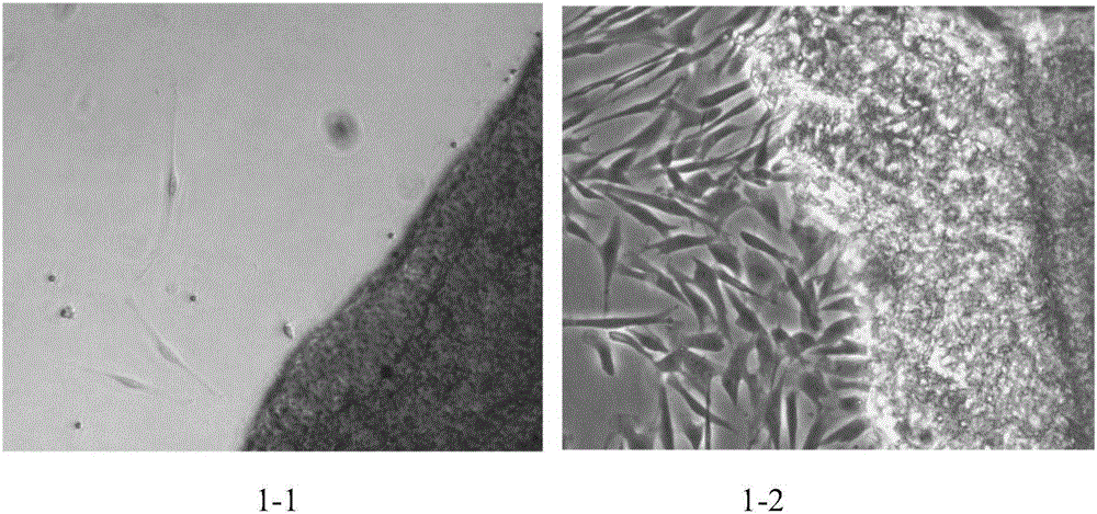
[0040] Example 1 Isolation of Placental Villi Plate Mesenchymal Stem Cells

[0041] (1) Sign an informed consent form with the donor, collect the full-term placenta, and process it as follows within 72 hours: Use sterile hemostatic forceps to peel off the amniotic membrane covering the outside of the villi plate; use sterile surgical scissors to cut a sufficient amount of the placenta villi plate organize;

[0042] (2) Use sterile tissue forceps to remove blood vessels and villi attached to the placenta villi;

[0043](3) Wash the placental villi plate obtained in step (2) with sterile physiological saline until there is no blood color;

[0044] (4) Use sterile surgical scissors to cut the cleaned villi plate tissue into 2mm 2 The tissue block was washed 2-5 times with sterile saline, avoiding the use of antibiotics during the cleaning process;

[0045] (5) Collect the tissue pieces in step (4), inoculate 2 g of tissue pieces into a T175 culture flask, add 15 mL of complete...

Embodiment 2

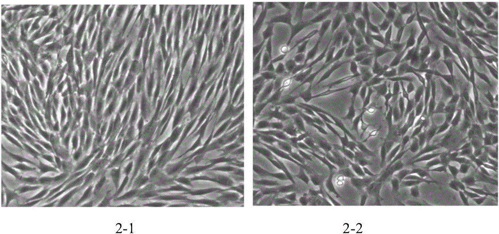
[0047] Example 2 Subculture and cryopreservation of placental villi plate mesenchymal stem cells

[0048] After the isolated placental villi plate mesenchymal stem cells were cultured until the cell confluency reached 70%-80%, trypLE TM Select (Gibco) was digested and centrifuged at 2000r / min for 3min, then the cells were collected, the cell viability was detected, and the cell count was performed according to 1×10 4 cells / cm 2 Density inoculation in T175 culture flasks, and adding 20mL complete medium for subculture, and then subculture the cells at a density of 2×10 6 cells / mL were resuspended in freezing medium (composition and mass fraction: 80% serum-free medium (Cellgenix), 13% human albumin (Sigma), 7% dimethyl sulfone (Origen)), and frozen The volume is 1mL, and then it is cooled to -80°C by a program and placed in a liquid nitrogen tank for long-term storage. See the results of subculture figure 2 .

Embodiment 3

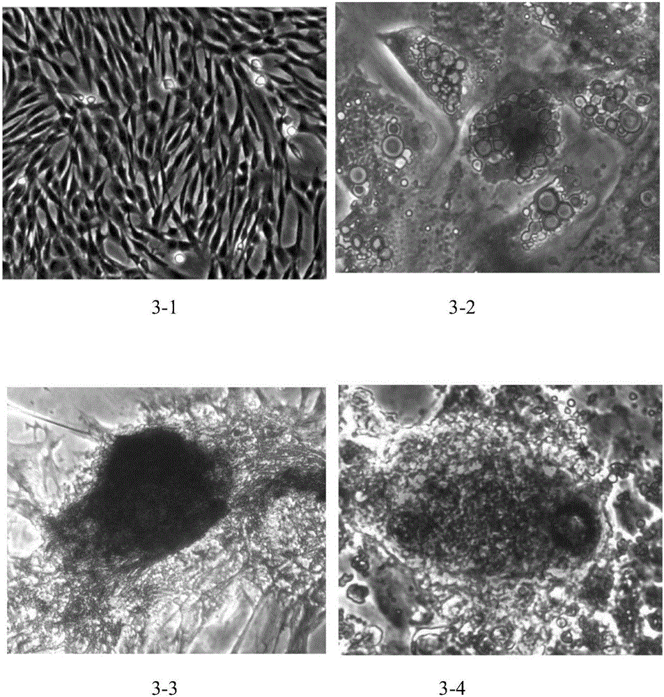
[0049] Example 3 Identification of Biological Characteristics of Placental Villi Plate Mesenchymal Stem Cells

[0050] 1. Identification of multi-lineage differentiation potential

[0051] The mesenchymal stem cells from P3 generation placental villi plate were inoculated in a six-well plate, and each well was inoculated with 1×10 5 Add 2 mL of complete medium to each well, set 3 replicates for each sample, replace the medium every two days, and add osteogenic, adipogenic, and chondrogenic induction medium (Cyagen) when the cell confluence is 70%. ) each 2mL, with the addition of complete medium as a blank control, the induction medium was replaced every 2-3 days, after continuous culture for 2-3 weeks, the induction medium was removed and washed once with PBS, and 2mL of paraformaldehyde solution (4% , v / v) fixed for 30min, then removed the paraformaldehyde solution and washed twice with PBS, stained with staining solution, the staining time for osteogenic induction and chon...


PUM


Abstract

The invention discloses placental villus plate mesenchymal stem cells. A clinical preparation method of the placental villus plate mesenchymal stem cells comprises the following steps: (1) acquiring a placental villus plate; (2) removing blood vessels and villus tissues; (3) cleaning the placental villus plate by virtue of sterile physiological saline until the placental villus plate has no blood color; (4) cutting villus plate tissues into 1-5mm[2], and cleaning the cut villus plate tissues by virtue of physiological saline; (5) inoculating tissue blocks into a complete medium, and cultivating the tissue blocks in a moist incubator which is at 37 DEG C and contains 5% of CO2; (6) changing the medium once every 6-11 days, and digesting the cells by virtue of TrypLE<TM> Select when a cell fusion degree is 70-80%; (7) conducting subculture; (8) conducting detection; (9) conducting cryopreservation; and (10) building a data base. According to the clinical preparation method disclosed by the invention, the placental villus plate is taken as a material, such operations as sorting, soaking, filtration and gradient centrifugation are not needed in an extracting process, so that many cells can be obtained by a relatively few of generation times; therefore, cost consumption and separation time can be greatly reduced, and the yield and purity of the mesenchymal stem cells can be improved.

Description

technical field [0001] The invention relates to the technical field of preparation of mesenchymal stem cells, in particular to a placental villi plate mesenchymal stem cell and a clinical preparation method. Background technique [0002] Mesenchymal stem cells (MSCs) are a type of adult stem cells with multi-directional differentiation potential, which can differentiate into cells of various tissue origins under specific induction conditions, such as adipocytes, osteoblasts, and chondrocytes. and neuroblasts, etc. Mesenchymal stem cells not only have multidirectional differentiation potential, but also have various biological characteristics such as self-renewal, homing, immune regulation, hematopoietic support, secretion of various cytokines, and promotion of stem cell transplantation. They are involved in tissue damage repair and tissue regeneration. important cellular components. Currently, mesenchymal stem cells can be extracted from various adult tissues (such as bone...

Claims


Application Information

Patent Timeline
IPC IPC(8): C12N5/0775
CPCC12N5/0668C12N2509/10
Inventor 李俊
Owner 四川华皓生物科技有限公司
Why Eureka
Try Eureka