Preparation method of amphoteric ion modified ultra-fine iron oxide particles

A technology of iron oxide particles and zwitterions, which can be used in preparations for in vivo tests, pharmaceutical formulations, nuclear magnetic resonance/magnetic resonance imaging contrast agents, etc. It can solve the problems of not finding MRI diagnostic applications and achieve the realization of magnetic resonance blood pools The effect of imaging, easy operation, and increased half-life of blood circulation

Inactive Publication Date: 2017-02-22
DONGHUA UNIV +1
View PDF1 Cites 11 Cited by
  • Summary
  • Abstract
  • Description
  • Claims
  • Application Information

AI Technical Summary

Problems solved by technology

[0004] Searching the literature at home and abroad, there is no relevant report on the zwitterionic L-cyste

Method used


Image

  • Preparation method of amphoteric ion modified ultra-fine iron oxide particles
  • Preparation method of amphoteric ion modified ultra-fine iron oxide particles
  • Preparation method of amphoteric ion modified ultra-fine iron oxide particles

Examples


Embodiment 1

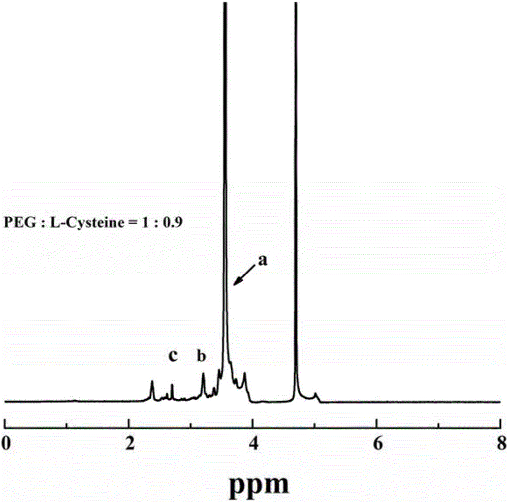
[0049] 1.09g of ferric chloride hexahydrate was dissolved in 40mL of diethylene glycol (also known as diethylene glycol, DEG), and then 0.47g of sodium citrate (Na 3 Cit) was dissolved in the above solution, and stirred at 80° C. for 1 hour under an air atmosphere. After the sodium citrate was completely dissolved, 1.312 g of anhydrous sodium acetate was added to the above solution, and the stirring was continued until the sodium acetate powder was completely dissolved, and then Transfer the solution to a 50mL autoclave, and react at 200°C for 4 hours; after the reaction, cool to room temperature naturally, transfer the product to a 50mL centrifuge tube and centrifuge at 8500rpm for 15 minutes, discard the supernatant, and return to the solution with absolute ethanol. Dissolve, centrifuge at 8500rpm for 15 minutes, repeat the operation 3 times, and then dry the precipitate at 60°C to obtain ultra-small ferric oxide nanoparticles, that is, ultra-small iron oxide nanoparticles st...

Embodiment 2

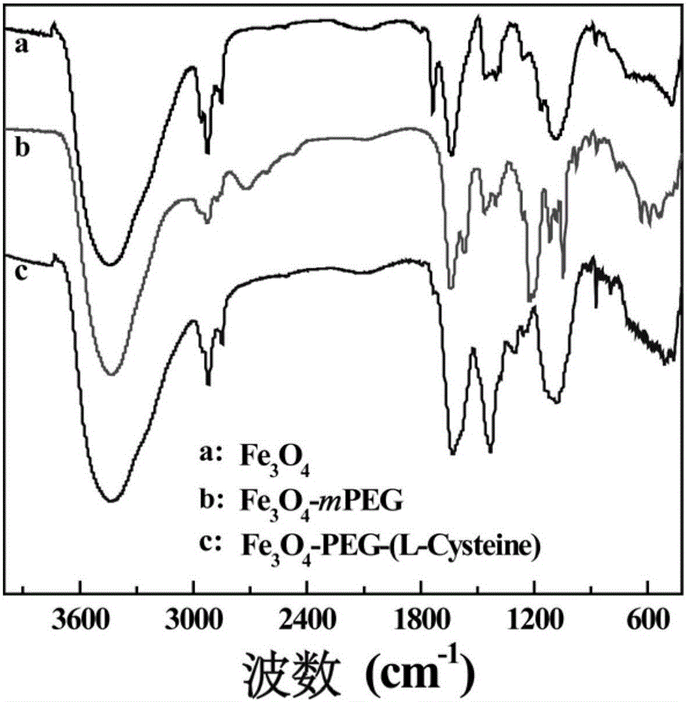
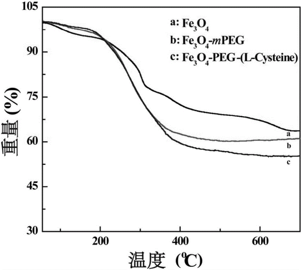
[0056] Get the Fe prepared by embodiment 1 respectively 3 o 4 , Fe 3 o 4 -PEG-(L-Cysteine) nanoparticles and the Fe prepared in Comparative Example 1 3 o 4 - Dissolve 2 mg of mPEG nanoparticles in ultrapure water to obtain a nanoparticle suspension, which is homogenized by ultrasound, and the surface potential and hydrated particle size are measured. Experimental result shows (table 1), the Fe that prepares 3 o 4 , Fe 3 o 4 -PEG-(L-Cysteine) and Fe 3 o 4 - The surface potentials of mPEG nanoparticles were -39.7, -15.7 and -16.4mV; the hydrated particle sizes were 30.9, 116.2 and 93.6nm, respectively. From the experimental results, the ultra-small iron oxide nanoparticles in the modification of NH 2 -PEG-(L-Cysteine) and mPEG-NH 2 After that, the surface potential increased and the hydrodynamic diameter increased, which was mainly caused by the surface modification of PEG or PEG / L-Cysteine. The changes of surface potential and hydrated particle size indicated that ...

Embodiment 3

[0061] In most cases, the route of administration of the contrast agent enters the body through intravenous injection. Therefore, the contrast agent will inevitably come into direct contact with the blood, and whether the intervention of the contrast agent will cause hemolysis or other adverse symptoms has become one of the important factors that researchers have to consider. In order to ensure that the nanoparticles prepared by the present invention can be safely used for in vivo bioimaging diagnosis, the prepared Fe 3 o 4 -PEG-(L-Cysteine) nanoparticles and control material Fe 3 o 4 - Hemocompatibility of mPEG. Calculate and weigh Fe according to the iron concentration calculation of two kinds of materials measured in embodiment 2 3 o 4 -PEG-(L-Cysteine) nanoparticles (embodiment 1) and contrast material Fe 3 o 4 -Two kinds of nanoparticles with 1 mg total iron content in mPEG (comparative example 1) were respectively dispersed in PBS to prepare a concentration of 1 m...


PUM

PropertyMeasurementUnit
Particle sizeaaaaaaaaaa
Hydrated particle sizeaaaaaaaaaa

Abstract

The invention relates to a preparation method of amphoteric ion modified ultra-fine iron oxide particles. The preparation method comprises the following steps: dissolving trivalent ferric salt in solvents, adding sodium citrate, stirring, adding anhydrous sodium acetate, stirring, reacting for 3 to 4 hours at the temperature of 190 to 200 DEG C, cooling, carrying out centrifugation and drying to obtain ultra-fine Fe3O4 nanoparticles, then dispersing the nanoparticles in ultrapure water, performing ultrasound, and EDC and NHS activation, dropwise adding to the ultrapure aqueous solution of Mal-PEG-NH2, reacting for 60 to 72 hours to obtain an aqueous solution of Fe3O4-PEG-Mal, and then dropwise adding the ultrapure aqueous solution of L-cysteine, reacting for 60 to 72 hours, dialyzing and freeze-drying to obtain the amphoteric ion modified ultra-fine iron oxide particles. The method disclosed by the invention is simple, and the prepared nanoparticles are long in half-life periods of blood in mouse bodies, and can realize blood pool angiography in animal levels and enhance imaging for subcutaneous transplanted tumors T1 of HeLa cells, so that the preparation method of the mphoteric ion modified ultra-fine iron oxide particles has potentials of industrialization and commercialization.

Description

technical field [0001] The invention belongs to the field of preparation of magnetic resonance imaging (MRI) contrast agents, in particular to a preparation method of zwitterion-modified ultra-small iron oxide particles. Background technique [0002] Malignant tumors have always been the number one killer of human life, with the characteristics of high mortality, difficult treatment and rapid deterioration. Therefore, early diagnosis and specific treatment of tumors are particularly important. Currently, tumor detection methods mainly include ultrasound imaging, CT imaging, nuclear medicine (PET or SPECT) imaging, and magnetic resonance imaging (MRI). With the development of magnetic resonance technology, the scanning time is gradually shortened, the resolution is gradually improved, and the detection of small lesions is more accurate, which also makes magnetic resonance imaging technology a new disease detection method developed in recent years. In order to improve the se...

Claims


Application Information

Patent Timeline
IPC IPC(8): A61K49/18A61K49/08A61P35/00
CPCA61K49/08A61K49/1836A61K49/186
Inventor 史向阳马丹罗宇王悍陈静文
Owner DONGHUA UNIV
Why Eureka
Try Eureka