Skull photon transparentizing treatment method and application thereof

A transparent processing and processing method technology, applied in the field of biomedical optical imaging, can solve the problems of skull turbidity, changing the physical and chemical properties of the cortex, and difficult surgical operations, so as to improve imaging contrast and signal-to-noise ratio, reduce internal tissue scattering, and enhance penetration. The effect of depth

Active Publication Date: 2017-06-30
HUAZHONG UNIV OF SCI & TECH
View PDF6 Cites 0 Cited by
  • Summary
  • Abstract
  • Description
  • Claims
  • Application Information

AI Technical Summary

Problems solved by technology

[0003] However, due to the high scattering properties of the skull above the cortex, the skull becomes very turbid, and it is difficult to focus photons in deep brain imaging. To solve this problem, researchers have mainly developed different cranial windows. The first is to use craniectomy During the operation, part of the skull was completely removed and the exposed meninges were covered with a cover glass. Due to the inevitable exposure of the brain, the physical and chemical properties of the cortex were changed, causing different degrees of inflammatory reactions, which affected the reliability and

Method used


Image

  • Skull photon transparentizing treatment method and application thereof
  • Skull photon transparentizing treatment method and application thereof
  • Skull photon transparentizing treatment method and application thereof

Examples


Example Embodiment

[0039] Example 1

[0040] The biological tissue of the embodiment of the present invention is taken from the isolated skull of a C57 mouse, and the intact skull is processed by the skull optical transparency treatment method of the embodiment 1 of the present invention, which specifically includes the following steps:

[0041] First, drip collagenase with a concentration of 2%-10% onto the skull of the mouse and apply evenly (1.5-2ml / cm 2 ), the treatment time is 5-7min; then wash the mouse skull with 80% absolute ethanol for 2-3min; then use 60%-80% fructose to smear the cleaned skull evenly, treatment 3- 5min; Finally, use a cyanoacrylic film to separate the above reagents from water, and place them under a water mirror for imaging to improve the matching degree of refractive index.
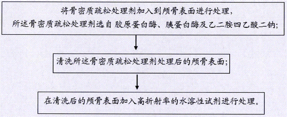
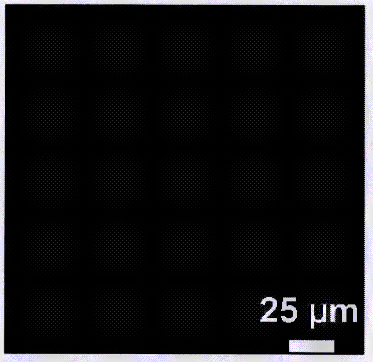
[0042] Cover the skull after optical transparency treatment in Example 1 of the present invention on the encapsulated 1.9μm diameter fluorescent bead solution, use a two-photon fluorescence microscop...

Example Embodiment

[0044] Example 2

[0045] Cut the scalp of the transgenic Thy-1-YFP-H line mouse to expose the skull of about 1cm×1cm square. The above-mentioned skull is processed by the optical transparency treatment method in Example 2 of the present invention, which specifically includes the following steps:

[0046] First, drop ethylenediaminetetraacetic acid disodium with a concentration of 2%-10% onto the skull of the mouse, and smear it evenly (0.5-0.8ml / cm 2 ), the treatment time is 5-7min; then wash the mouse skull with water for 2-3min; then use 60%-80% sorbitol to smear the cleaned skull evenly and treat it for 3-5min; finally use cyanoacrylic acid The film separates the water and the light transparent agent and places it under a water mirror for imaging to improve the matching degree of the refractive index.

[0047] Figure 3a-3e Fluorescence contrast images of cortical dendrites and dendritic spines taken before and after the light-transparency treatment of the mouse skull in Example...

Example Embodiment

[0048] Example 3

[0049] The wild-type C57 mouse scalp is cut open to expose the skull of about 1cm×1cm square, and the above-mentioned skull is processed by the light transparent processing method in Example 3 of the present invention, which specifically includes the following steps:

[0050] First, add trypsin with a concentration of 2%-10% to the skull of the mouse and apply evenly (0.5-0.8ml / cm 2 ), the treatment time is 5-7min; then wash the skull with 90% absolute ethanol for 2-3min; then use 60%-80% sucrose to smear the cleaned skull evenly and treat it for 3-5min; Finally, a cyanoacrylic film is used to separate the water and the light transparent agent, and placed under a water mirror for imaging to improve the matching degree of the refractive index.

[0051] Figure 4a-4b The contrast images of the cortical blood vessels taken before and after the light transparent treatment of the mouse skull in Example 3 of the present invention under living conditions are given. It c...


PUM


Abstract

The invention provides a skull photon transparentizing treatment method and application thereof. The treatment method comprises the following steps that a compact bone substance loosening agent is added to the skull surface for treatment, and the compact bone substance loosening agent is selected from collagenase, trypsin and disodium edetate dehydrate; the skull surface treated with the compact bone substance loosening agent is cleaned; a high-refraction-index water-soluble reagent is added to the cleaned skull surface for treatment, and the skull with the relatively uniform refraction index is obtained. The skull structure treated through the method becomes transparent, and can be used for living body two-photon microimaging, and high-resolution imaging of cortex nerves and microvascular structures is achieved.

Description

technical field [0001] The invention belongs to the technical field of biomedical optical imaging, and in particular relates to a skull optical transparency processing method and an application thereof. Background technique [0002] The long-term observation and manipulation of cortical cells is of great significance for the study of neurons, glial cells and microvasculature. With the development of transgenic technology and two-photon microscopic imaging, scientists can conduct research on the cortex at the level of living mammals. Repeated imaging over time to understand the complex environment of the nervous system. Especially to track the fate of individual synapses, and then study the regulation of sensory experience, learning and memory on the dynamic changes of dendritic spines. [0003] However, due to the high scattering properties of the skull above the cortex, the skull becomes very turbid, and it is difficult to focus photons in deep brain imaging. To solve this...

Claims


Application Information

Patent Timeline
IPC IPC(8): A61D7/00A61B5/00
Inventor 赵延洁
Owner HUAZHONG UNIV OF SCI & TECH
Why Eureka
Try Eureka