
Preparation method and application of fluorine-19 magnetic resonance imaging probe for sulfydryl detection

A magnetic resonance and sulfhydryl technology, applied in the field of nanoprobes, can solve the problems of background interference, poor imaging effect, affecting the diagnosis results, etc., and achieve the effect of easy analysis and processing and high selectivity.

Active Publication Date: 2018-02-13
INST OF BIOMEDICAL ENG CHINESE ACAD OF MEDICAL SCI
View PDF5 Cites 1 Cited by
  • Summary
  • Abstract
  • Description
  • Claims
  • Application Information

AI Technical Summary

Problems solved by technology

However, clinical practice has found that due to the wide distribution of water molecules in the body, there is strong background interference. In this case, the imaging effect obtained is very poor, which affects the judgment of the diagnosis result.

Method used


Image

  • Preparation method and application of fluorine-19 magnetic resonance imaging probe for sulfydryl detection
  • Preparation method and application of fluorine-19 magnetic resonance imaging probe for sulfydryl detection
  • Preparation method and application of fluorine-19 magnetic resonance imaging probe for sulfydryl detection

Examples


Embodiment 1

[0025] Synthesis of embodiment 1 polymer P4

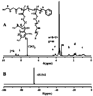
[0026] Add the reversible addition-fragmentation chain transfer polymerization (RAFT) chain transfer agent phenyl-(2-hydroxyethyl) thiocarbonate (12.2 mg, 0.1 mM) and polyethylene glycol monomethyl ether in sequence in the shlenk reaction tube Methyl methacrylate (mPEGMA) (500 mg, 1 mM) and 2-((2,4-dinitro-N-(3,3,3-trifluoropropyl)phenylsulfonamide) ethyl methacrylate Ester (AMA-DNBSF) (455 mg, 1 mM), initiator (AIBN) (1.64 mg, 0.01 mM) and 3 mL solvent dimethylformamide (DMF), after three cycles of vacuum / argon, Seal the reaction tube and react in an oil bath at 68-72°C for 24 h. After the reaction, add DMF to dissolve it, put it into a dialysis bag (molecular weight cut-off 3500 Da), dialyze with deionized water for 72 h, and replace it every 12 h Dialysate. Afterwards, lyophilization obtains PEDF polymkeric substance. Utilize proton nuclear magnetic spectrum and fluorine spectrum to carry out characterization to P4 polymkeric s...

Embodiment 2

[0030] Example 2 Preparation method of nanoprobe N4

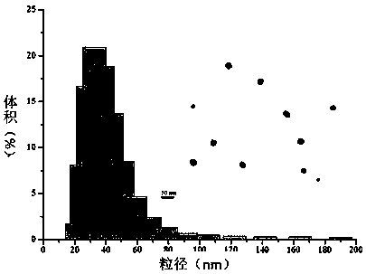
[0031] Weigh 20 mg of PEDF polymer and directly disperse it in 10 mL of 0.01 M phosphate buffer solution with pH=7.4 under the action of ultrasound (25 °C, 50 Hz, 5 min) to obtain N4 nanoparticles with a concentration of 2 mg / mL. The particle size and shape of nanoparticles were detected by laser particle size analyzer and transmission electron microscope, and the detection results are shown in the attached figure 2 As shown, the nanoparticles prepared in this example have a particle size of 45 nm, a particle size distribution of 0.12, and an obvious core-shell structure.

[0032] According to the preparation method in Example 2, nanoparticles composed of other polymers can be prepared.

Embodiment 3

[0033] Example 3 Nanoprobe N4 19 F NMR signal changes with reaction time

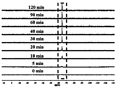
[0034] Take 5 mL of 2 mg / mL N4 nanoprobe solution, add cysteine ​​(Cys) mother solution under argon protection conditions, so that the final concentration of Cys is 5 mg / mL, at the set time point, take 300 µL, add 100 µL heavy water for field lock, use nuclear magnetic resonance to analyze the 19 F NMR signal was detected. as attached image 3 As shown, the nanoprobe can respond rapidly to sulfhydryl groups, in the presence of cysteine, within 30 min 19 The F NMR signal can be significantly enhanced.


PUM


Abstract

The invention relates to a preparation method and application of a fluorine-19 magnetic resonance imaging probe for sulfydryl detection. The structure and composition of the probe is as shown in the specification, wherein x is an integer from 5 to 50, and y is an integer from 5 to 50. The probe is a novel <19>F magnetic resonance imaging nano probe which uses redox response to activate amino protonation and is used for detecting in-vivo sulfydryl. The nano probe has the advantages that the nano probe is high in selectivity to sulfydryl compounds, and a detection signal can be easily analyzed and processed; meanwhile, the nano probe is high in sensitivity, simple to operate, small in tissue damage, high in resolution and the like, and an important means is provided for the real-time and in-situ visual monitoring of the in-vivo sulfydryl compounds.

Description

technical field [0001] The invention relates to a preparation method and application of a fluorine-19 magnetic resonance imaging probe for sulfhydryl detection, in particular to a polymer nanoprobe suitable for highly sensitive and highly selective sulfhydryl detection, using fluorine-19 magnetic resonance imaging ( 19 F MRI) detects sulfhydryl groups in cells or in vivo levels, and belongs to nanoprobes in the field of bioanalysis technology. Background technique [0002] In recent years, with the rapid development of life science and medical science, the detection of active substances in cells has gradually attracted widespread attention. Cysteine ​​(Cys), homocysteine ​​(Hcy) and glutathione (GSH) and other active sulfhydryl small molecules in organisms play an important role in human physiological and pathological processes. Although they are very similar in structure, their biological functions are completely different. Cys is the only natural essential amino acid co...

Claims


Application Information

Patent Timeline
Patent Type & Authority Applications(China)
IPC IPC(8): A61K51/06A61K49/12A61K101/02
CPCA61K49/126A61K51/06
Inventor 黄平升王伟伟秦怡博孔德领
Owner INST OF BIOMEDICAL ENG CHINESE ACAD OF MEDICAL SCI
Learn More