
Quantitative method and application for detection of protein drugs in vivo based on the specific separation-amplification lc-ms of tagged small molecules

An LC-MS, protein drug technology, applied in the field of medicine, can solve the problems that the detection sensitivity cannot meet the detection of low-concentration protein drugs, the proteolysis time is long, and the operation requirements are high, so as to overcome the bottleneck of protein drug concentration, improve the detection sensitivity, good repeatability

Active Publication Date: 2020-12-15
南京杰宁医药科技有限公司
View PDF0 Cites 0 Cited by
  • Summary
  • Abstract
  • Description
  • Claims
  • Application Information

AI Technical Summary

Problems solved by technology

At present, the proteolysis method is used to enzymatically hydrolyze protein drugs and select several characteristic peptides for quantitative analysis. However, this technology only converts protein drugs into characteristic fragments with slightly higher ion efficiency than protein drugs for analysis. The detection sensitivity achieved still cannot meet the detection of low concentration protein drugs
At the same time, the process of proteolysis is time-consuming and complicated, and requires high operation, which is easy to introduce large errors.

Method used


Image

  • Quantitative method and application for detection of protein drugs in vivo based on the specific separation-amplification lc-ms of tagged small molecules
  • Quantitative method and application for detection of protein drugs in vivo based on the specific separation-amplification lc-ms of tagged small molecules
  • Quantitative method and application for detection of protein drugs in vivo based on the specific separation-amplification lc-ms of tagged small molecules

Examples


Embodiment 1

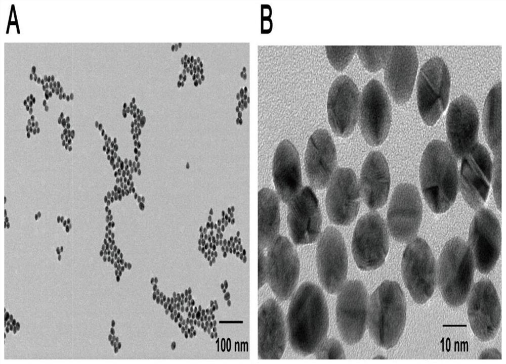
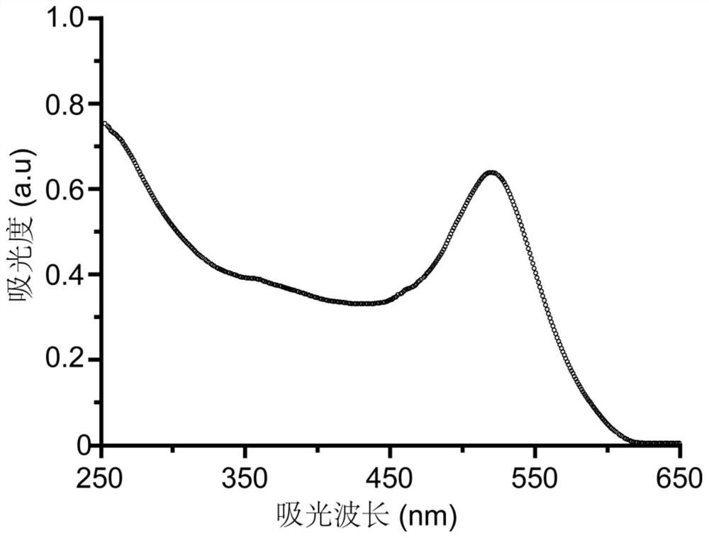
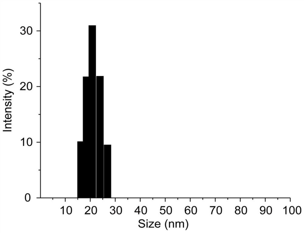
[0042] Preparation of functionalized gold nanoprobes:

[0043]Weigh an appropriate amount of chloroauric acid and dissolve it with ultrapure water to prepare 100mL of 1mM chloroauric acid aqueous solution, transfer it to a round bottom flask and heat to boiling, then quickly add 10mL of the prepared 38.8mM sodium citrate aqueous solution, and continue to boil and reflux for 15 minutes, remove the heat source, continue to reflux, stir and cool to room temperature to obtain a gold nanoparticle solution. Take out 1 mL of the gold nanoparticle solution, add 60 μL of 50 μg / mL adenosine aqueous solution, shake and mix at room temperature for 30 minutes to obtain the gold nanoparticle / adenosine complex. Activate the aptamer with TCEP 50 times that of the thiol aptamer at room temperature for 1 hour, pipette the nano-gold / adenosine complex into the activated thiol aptamer solution, shake it at room temperature in the dark for 16 hours, and add it dropwise 10 μL of Tris-acetic acid bu...

Embodiment 2

[0045] Preparation of functionalized magnetic bead probes:

[0046] Weigh silicon amide-coated magnetic beads, ultrasonically disperse and wash them several times with ultrapure water, and finally disperse in 10 mM PBS buffer solution (pH, 8.0), add glutaraldehyde aqueous solution to the system, and shake at room temperature for 1.5 hours. Apply an external magnetic field to remove the supernatant, add 3×SSC buffer to redisperse, add excess aminated aptamer solution, shake well at room temperature and react overnight. Add sodium borohydride aqueous solution to react for 0.5 hours, wash with 3×SSC buffer solution, 0.2% SDS aqueous solution, and finally wash with ultrapure water for 3 times, remove the supernatant with an external magnetic field, and disperse in 10mM Tris-HCl buffer solution (pH, 7.6).

Embodiment 3

[0048] The formation of the double-wrapped sandwich structure:

[0049] The functionalized magnetic bead probe was repeatedly washed 3 times with PBS buffer (10mM, pH, 7.4), and the supernatant was removed by an external magnetic field, and then redispersed in a K-containing solution. + , Mg 2+ In the activation buffer system, after activation at room temperature for 1 hour, add the sample to be tested containing thrombin, place it at 37°C for 2 hours, mix and react at a constant temperature, remove the supernatant, and prepare the sample containing K + , Mg 2+ and 0.005% Tween-20 (w / v) washing buffer to wash gently for 3 times, and remove the supernatant. After the functionalized gold nanoprobe was centrifuged to remove the supernatant, it was redispersed in a solution containing K + , Mg 2+ In the activation buffer system, and transferred to the magnetic bead probe sample after removing the supernatant, placed at 37°C for a constant temperature mixing reaction for 2 hour...


PUM


Abstract

The invention relates to an analytical method applied to high specificity and high sensitivity detection of a trace protein drug in a biological sample. Taking thrombin as a study example, constructedfunctionalized superparamagnetic nanoparticles specifically capture the thrombin in the sample, and further form a sandwich complex with a dual functionalized nanogold probe of which the surface simultaneously modifies a large amount of label small molecules and an aptamer of specific binding protein; on the basis of the sandwich structure, transformation of a detection target is realized, namely, the label small molecules instead of the protein drug are used for further mass spectrometric detection, so that the difficulties of low response for direct mass spectrometric detection of protein macromolecules and the like are overcome; moreover, by constructing the sandwich structure, the large amount of label small molecules proportioned to to-be-detected target protein are introduced and further the amplification of a detection signal is realized; besides, a novel method which is sensitive and efficient, does not need protease hydrolysis and directly measures the concentration of the protein drug by the LC-MS / MS by combining high specificity of the sandwich structure with high sensitivity of LC-MS / MS. A preparation method disclosed by the invention has the advantages of simplicity,simplicity and convenience in operation, economy, effectiveness, quickness and sensitiveness, and starts a new stage for LC-MS / MS detection of the protein drug in the body.

Description

technical field [0001] The invention belongs to the field of medicine, and specifically relates to a quantitative method for detecting protein drugs in vivo based on LC-MS / MS of biological barcode exclusive separation-amplification-no proteolysis pretreatment. Background technique [0002] Recently, more and more protein drugs have been developed and applied to the diagnosis and treatment of diseases, showing obvious advantages in the development of medicine, and have become one of the rapidly developing hot spots in the research of new drugs today. In order to promote the development and biological application of protein drugs, it is an important research link to correctly evaluate the in vivo pharmacokinetics of protein drugs. However, protein drugs have problems such as high biological activity, strong targeting, small dosage required, low concentration in vivo, and susceptibility to interference by endogenous substances, which pose great challenges to traditional analysi...

Claims


Application Information

Patent Timeline
Patent Type & Authority Patents(China)
IPC IPC(8): G01N30/02
CPCG01N30/02G01N2030/027
Inventor 杨文丁黎
Owner 南京杰宁医药科技有限公司
Learn More