Protein chip for simultaneously detecting four bladder cancer markers in urine
A protein chip, bladder cancer technology, applied in measurement devices, biological tests, material inspection products, etc., can solve the problems of a large proportion of false negatives, unsuitable for rapid detection, specificity and sensitivity need to be improved, and achieve a simple preparation process. , The effect of chip detection method is fast and cheap
- Summary
- Abstract
- Description
- Claims
- Application Information
AI Technical Summary
Problems solved by technology
Method used
Image
Examples
Embodiment 1
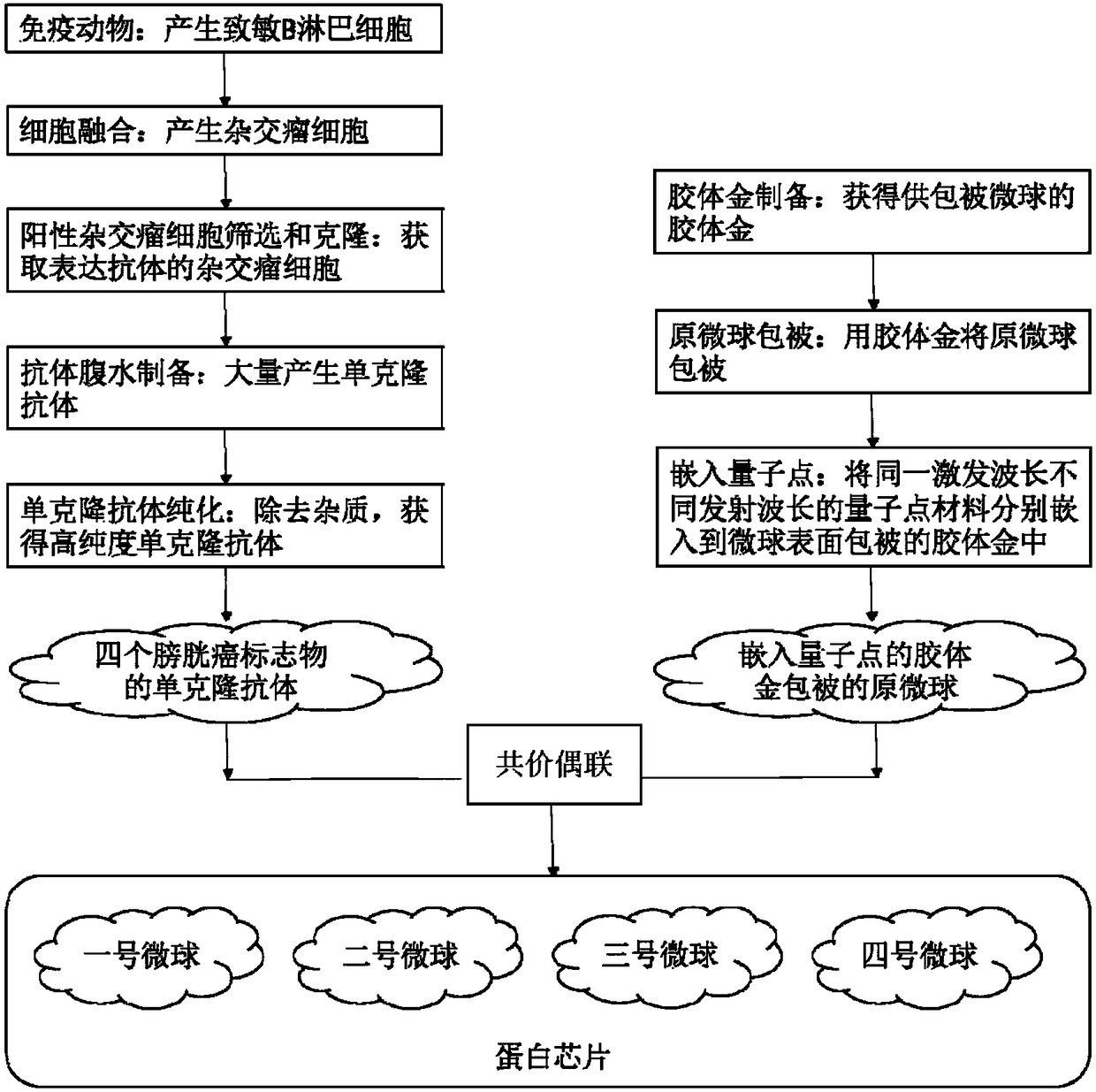
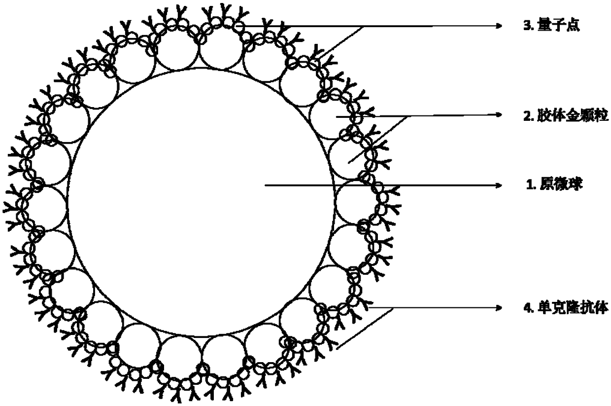
[0040] Example 1: Preparation of colloidal gold-coated microspheres and embedding of quantum dots:
[0041] 1. Preparation of colloidal gold
[0042] The colloidal gold particles prepared according to the following method in the present invention have a particle diameter of about 20 nm, and a gold concentration of about 0.1 mg / mL.
[0043] The specific preparation method is: first chloroauric acid (HAUC 14 ) into a 0.01% aqueous solution, take 100mL to 250mL beaker, pickle the beaker, rinse it with distilled water and siliconize it before use. After heating to boiling, add sodium citrate and observe the color change of the chloroauric acid aqueous solution under the boiling state, it can be seen that the chloroauric acid aqueous solution changes from light yellow to gray, then becomes black, then gradually transitions to red and remains stable. Change, the whole process is about 3 minutes. After the red aqueous solution is cooled to room temperature, restore to the origin...
Embodiment 2
[0049] Example 2: Preparation of monoclonal antibodies and coupling to colloidal gold-coated primary microspheres embedded in quantum dots:
[0050] 1. Cell fusion and preparation of hybridoma cells
[0051] Female Balb / c mice aged 6-8 weeks were selected, and recombinant proteins of NMP22, BTA, CD20, and TERT were injected intraperitoneally as antigens, so that the antigens could enter the peripheral immune organs through blood circulation or lymphatic circulation, and stimulate corresponding B lymphocyte clones. They activate, proliferate, and differentiate into primed B lymphocytes. After three days, adopt CO 2 The mice were killed by gas, and the spleen was removed under aseptic operation to prepare a spleen cell suspension. Take mouse myeloma cells and splenocytes in the logarithmic growth phase at a ratio of 5:1, mix them in a sterile and non-toxic environment in a water bath at 40°C, add 50% polyethylene glycol (PEG) as a fusion-promoting agent, and make Lymphocyte...
Embodiment 3
[0060] Example 3: The protein chip prepared by the present invention detects four bladder cancer markers:
[0061] 1. Acquisition of samples to be tested:
[0062] The protein chip prepared by the invention is suitable for detecting trace markers in urine. The mid-morning urine was collected, immediately transferred to the laboratory, stored at low temperature for future use, and the test was completed within 12 hours.
[0063] 2. Co-incubation of the protein chip and the sample to be tested:
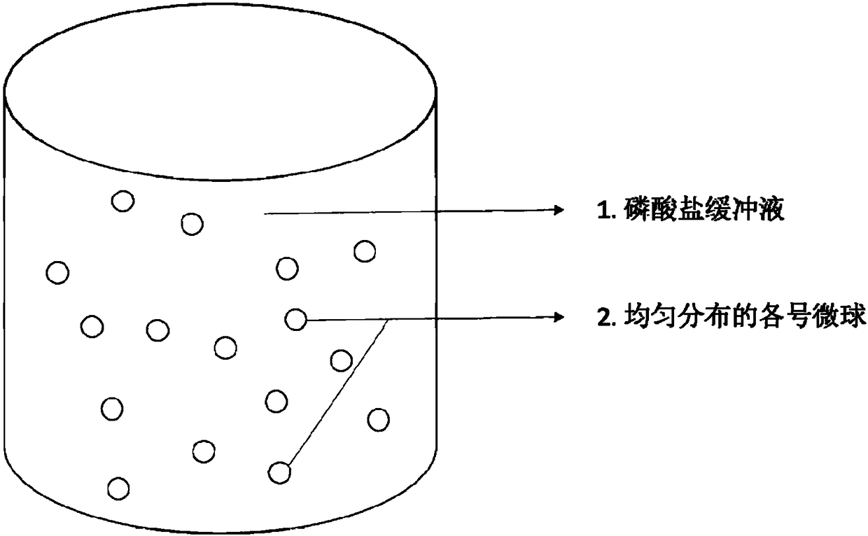
[0064] Take the protein chip stored in PBS buffer, and shake it sufficiently to form a microsphere suspension. Add the urine sample and the protein chip in the suspension state to the petri dish at one time, mix thoroughly, and react in a 37°C incubator in the dark for 45min. After taking out the culture dish, add an appropriate amount of microsphere suspension again, mix well, and react in a 37°C incubator in the dark for 30min.
[0065] 3. Detection of four bladder cancer marker...
PUM
| Property | Measurement | Unit |
|---|---|---|
| Particle size | aaaaa | aaaaa |
Abstract
Description
Claims
Application Information
 - R&D Engineer
- R&D Manager
- IP Professional
- Industry Leading Data Capabilities
- Powerful AI technology
- Patent DNA Extraction
Browse by: Latest US Patents, China's latest patents, Technical Efficacy Thesaurus, Application Domain, Technology Topic, Popular Technical Reports.
© 2024 PatSnap. All rights reserved.Legal|Privacy policy|Modern Slavery Act Transparency Statement|Sitemap|About US| Contact US: help@patsnap.com


