In-situ autologous bionic repair material for fat liquefaction wound as well as preparation method and application of in-situ autologous bionic repair material

A repair material and autologous technology, applied in the direction of prosthesis, pharmaceutical formulation, medical science, etc., can solve the problems of poor healing, unsatisfactory healing cycle and prognosis scar, infection, etc., achieve good liquid absorption and air permeability, reduce prognosis Poor function, the effect of promoting physiological repair

Inactive Publication Date: 2018-08-24
SHANGHAI SIXTH PEOPLES HOSPITAL
View PDF0 Cites 4 Cited by
  • Summary
  • Abstract
  • Description
  • Claims
  • Application Information

AI Technical Summary

Problems solved by technology

Fat liquefied wounds lead to increased chances of infection and poor healing. At present, incision drainage, repeated washing and dressing changes, physical therapy and other methods are mainly relied on. The treatment cycle is long and the burden on doctors and patients is heavy.
At present, there is no special medical product for fat liquefaction wounds. In recent years, the hemostatic effect of various biological materials on wounds has been clinically proven, such as chitosan, alginic acid, collagen, cellulose, fibrin, silk fibroin, polyurethane , silicone and other dressings, the above products can effectively absorb wound exudate, accelerate blood coagulation, and promote wound healing, but the healing cycle and prognosis of scars are not ideal, and cannot effectively meet clinical needs

Method used


Image

  • In-situ autologous bionic repair material for fat liquefaction wound as well as preparation method and application of in-situ autologous bionic repair material
  • In-situ autologous bionic repair material for fat liquefaction wound as well as preparation method and application of in-situ autologous bionic repair material
  • In-situ autologous bionic repair material for fat liquefaction wound as well as preparation method and application of in-situ autologous bionic repair material

Examples


Embodiment 1

[0037] Example 1 Preparation of in-situ autologous biomimetic repair material for fat liquefaction wounds

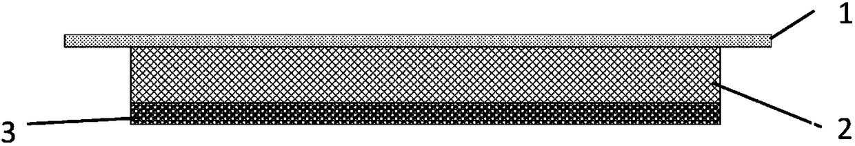
[0038](1) Preparation of hydrophilic polyurethane film: referring to the classic medical polyurethane film preparation process, first prepare a prepolymer from hydrophilic polyethylene glycol and isocyanate in the presence of a crosslinking agent and a catalyst, and then dissolve the prepolymer In ethyl acetate, cast into a film with a thickness of about 0.1mm, cut into suitable sizes, and set aside;

[0039] (2) Apply pressure-sensitive adhesive evenly on one side of the prepared polyurethane film, and set aside;

[0040] (3) Chitosan fiber is woven into a multi-layer gauze structure by textile technology. The liquid absorption of the fiber layer is about 17 times its own weight, and the water vapor transmission rate is about 3937g.m -2 24 hours -1 ; The biological fiber layer is cut into suitable sizes for subsequent use;

[0041] (4) The biological fiber layer is p...

Embodiment 2

[0043] Example 2 In situ autologous bionic repair material for fat liquefaction wounds

[0044] (1) with embodiment 1;

[0045] (2) with embodiment 1;

[0046] (3) Using textile technology to blend chitosan fiber and calcium alginate fiber into a multi-layer gauze structure, chitosan fiber accounts for 70%, calcium alginate fiber accounts for 30%; the liquid absorption of the fiber layer is about 15 times Self-weight, water vapor transmission rate is about 3720g.m -2 24 hours -1 ; The biological fiber layer is cut into suitable sizes for subsequent use;

[0047] (4) with embodiment 1;

[0048] (5) with embodiment 1.

Embodiment 3

[0049] Example 3 In situ autologous bionic repair material for fat liquefaction wound

[0050] (1) with embodiment 1;

[0051] (2) with embodiment 1;

[0052] (3) Chitosan fiber, calcium alginate fiber and oxidized cellulose fiber are blended into a multi-layer gauze structure by textile technology, chitosan fiber accounts for 40%, calcium alginate fiber and oxidized cellulose fiber each account for 30%; The liquid absorption capacity of the fiber layer is about 16 times its own weight, and the water vapor transmission rate is about 3877g.m -2 24 hours -1 . The biological fiber layer is cut into applicable size and set aside;

[0053] (4) with embodiment 1;

[0054] (5) with embodiment 1.


PUM

PropertyMeasurementUnit
thicknessaaaaaaaaaa
thicknessaaaaaaaaaa
thicknessaaaaaaaaaa

Abstract

The invention discloses an in-situ autologous bionic repair material for a fat liquefaction wound as well as a preparation method and an application of the in-situ autologous bionic repair material, and belongs to the field of medical materials. The in-situ autologous bionic repair material for the fat liquefaction wound comprises an autologous functional layer, a biological functional layer and asurface separation layer, wherein the autologous functional layer is PRP (platelet rich plasma) prepared after whole blood of a patient is collected; the biological functional layer contains one or more of chitosan fiber, alginic acid fiber, collagen fiber, oxidized cellulose fiber and gelatin fiber and is of a fibrous layer structure, and a 3D network structure of the biological functional layercan be applied to infiltration and immobilization of components in the autologous functional layer; the surface separation layer is a polyurethane semipermeable membrane. The in-situ autologous bionic repair material for the fat liquefaction wound is high in biosecurity and free of immune risk, can be used for treating the fat liquefaction wound which is difficult to heal or poor in healing, andcan also be used for wound care of special groups such as elderly cesarean parturient women.

Description

technical field [0001] The invention belongs to the technical field of biomedical materials, and in particular relates to an in-situ autologous bionic repair material for fat liquefaction wounds and a preparation method and application thereof. Background technique [0002] Fat liquefaction is one of the postoperative complications, and its essence is the outflow of lipid droplets caused by aseptic necrosis of adipocytes. Obesity is the main reason, diabetes, anemia, edema, high-frequency knife, etc. are also incentives. Since the liberalization of the two-child policy, the emergence of many elderly women has further increased the incidence of non-healing of fat liquefied wounds. Fat liquefaction wounds lead to increased chances of infection and poor healing. At present, incision drainage, repeated washing and dressing changes, physical therapy and other methods are mainly used. The treatment cycle is long and the burden on doctors and patients is heavy. At present, there ...

Claims


Application Information

Patent Timeline
Patent Type & Authority Applications(China)
IPC IPC(8): A61L27/40A61L27/36A61L27/20A61L27/24A61L27/22A61L27/18A61L27/50A61L27/54A61L27/56
CPCA61L27/3616A61L27/18A61L27/20A61L27/222A61L27/24A61L27/50A61L27/54A61L27/56A61L2300/404A61L2300/41A61L2300/412A61L2300/602A61L2400/04C08L5/08C08L5/04C08L1/04C08L75/04
Inventor 张长青位晓娟张伟
Owner SHANGHAI SIXTH PEOPLES HOSPITAL
Why Eureka
Try Eureka