Vibrio alginolyticus bacteriophage preparation and preparation method and application thereof
A technology of vibrio alginolyticus and bacteriophage, which is applied in botanical equipment and methods, applications, and medical raw materials derived from viruses/phages, etc. Easy to obtain, low price, small usage effect
- Summary
- Abstract
- Description
- Claims
- Application Information
AI Technical Summary
Problems solved by technology
Method used
Image
Examples
Embodiment 1
[0048] The separation and purification of embodiment 1 phage prepares phage
[0049] (1) Seawater treatment and phage enrichment
[0050] Vibrio alginolyticus E06333 was cultured overnight as the host bacteria for phage isolation. Seawater samples were centrifuged at 10,000 rpm for 10 min, and the supernatant was filtered through a 0.45 μm filter membrane to retain the filtrate. Add the filtrate to 2×LB medium at a volume ratio of 1:1, then add the overnight cultured E06333 bacterial solution at a volume ratio of 1%, culture at 30°C for 12 hours, and after centrifugation, filter the supernatant through 0.22 μm After membrane filtration, the phage enrichment solution was obtained.
[0051] (2) Phage purification
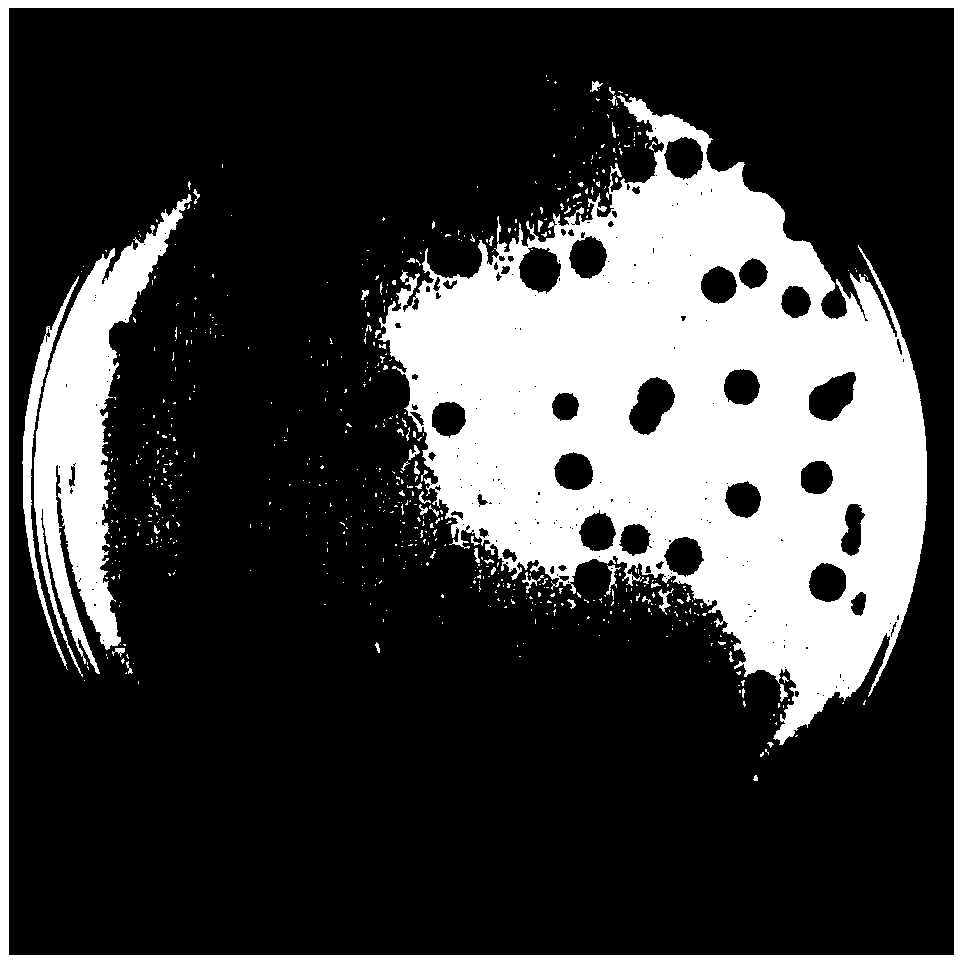
[0052] Using the double-layer plate method, the phage enrichment solution was serially diluted with sterilized SM buffer (0.05M Tris-HCl, pH 7.5), and 100 μL of Vibrio alginolyticus E06333 in the exponential phase was mixed with 100 μL of the phage serial dilution ...
Embodiment 2
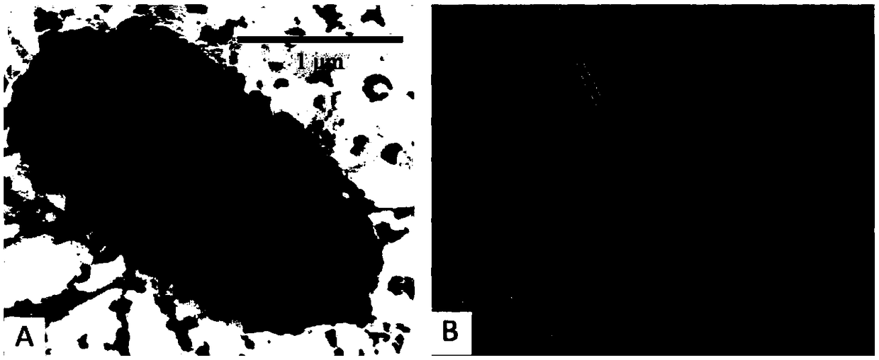
[0055] Example 2 Electron Microscopic Detection of Vibrio alginolyticus Phage Vp670
[0056] The Vibrio alginolyticus E06333 culture solution and the bacteriophage Vp670 preservation solution in the above-mentioned Example 1 were respectively dropped on the copper grid, and naturally precipitated for 2-3 minutes. Absorb excess liquid sideways with filter paper. Then stain with 2% phosphotungstic acid for 10 min. Then use filter paper to absorb excess dye solution from the side and air dry at room temperature. The morphology of Vibrio alginolyticus and phage Vp670 was observed and recorded by Hitachi transmission electron microscope.
[0057] Electron microscope observation results such as figure 2 As shown, it can be seen that the normal host bacteria cell structure is complete, and the texture inside the cell is uniform. The outer membrane layer of the Vibrio alginolyticus host bacteria infected by phage Vp670 disappeared, and a large light-colored area appeared inside, a...
Embodiment 3
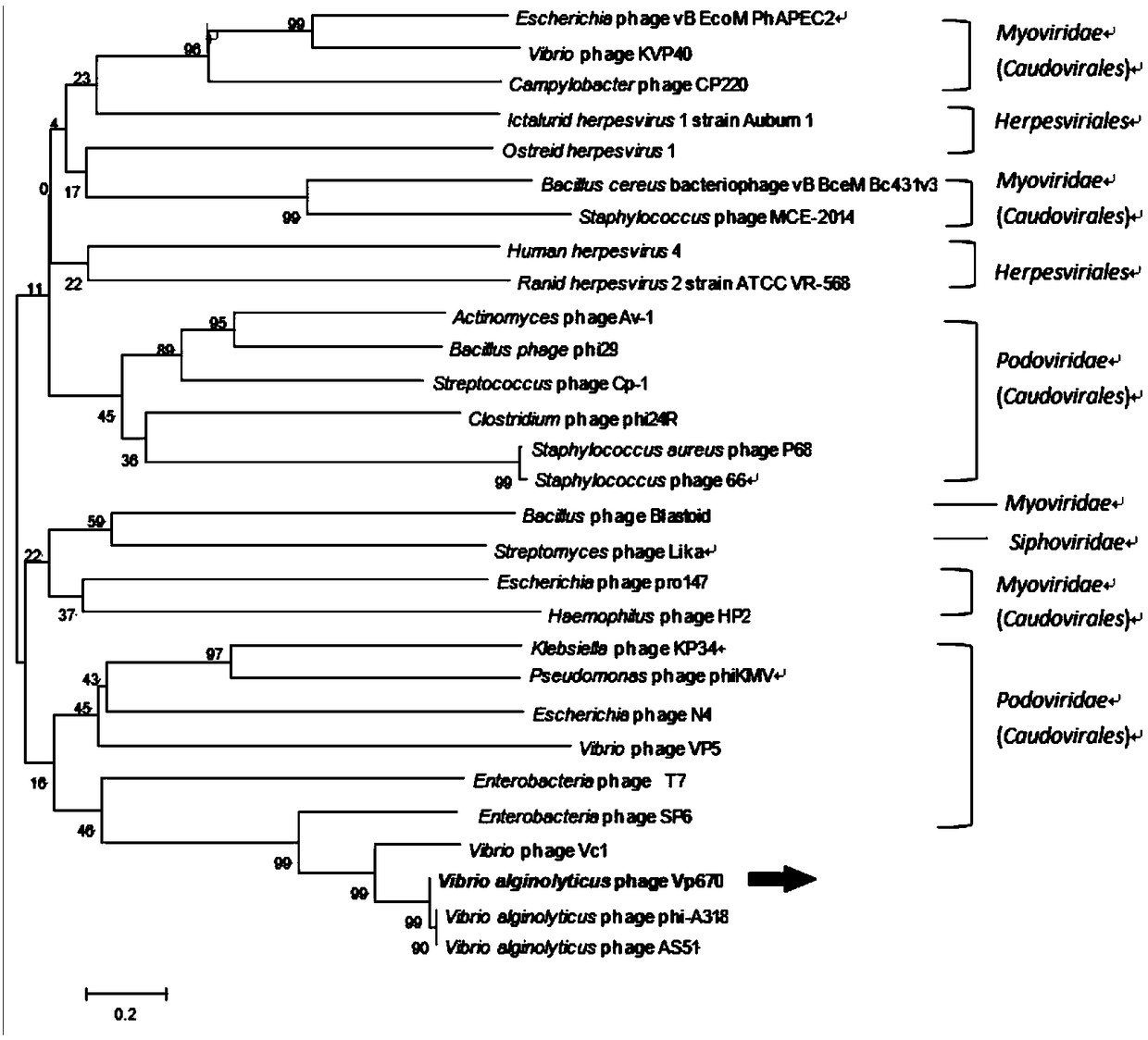
[0058] Example 3 Molecular identification of Vibrio alginolyticus phage Vp670
[0059] According to the genome sequence of phage Vp670 (GenBank: KY290756.1), its DNA polymerase (DNAP) sequence was found, and the phylogenetic tree was constructed using DNAP and DNAP sequences of similar phages, using the tree building tool MEGA 6.0. Phylogenetic Tree See image 3 . Depend on image 3 It can be seen that Vp670 is clustered with multiple phages of the Brachyphage family from different sources, and this branch is obviously different from the branches formed by other bacteriophage families. Therefore, combined with the electron microscope morphology observation, it is determined that the phage Vp670 belongs to the Brachyphage family.
PUM
 Abstract
Description
Claims
Application Information
 - R&D
- Intellectual Property
- Life Sciences
- Materials
- Tech Scout
- Unparalleled Data Quality
- Higher Quality Content
- 60% Fewer Hallucinations
Browse by: Latest US Patents, China's latest patents, Technical Efficacy Thesaurus, Application Domain, Technology Topic, Popular Technical Reports.
© 2025 PatSnap. All rights reserved.Legal|Privacy policy|Modern Slavery Act Transparency Statement|Sitemap|About US| Contact US: help@patsnap.com


