Tumor-specific cleavable pegized nanoparticles, preparation method and application

A tumor-specific, nanoparticle technology, applied in the field of tumor-specific cleavable PEGylated nanoparticles and their preparation, can solve problems such as prolonging circulation time, reducing drug targeted release and bioavailability, and achieving biocompatibility Good properties, strong drug loading capacity, and low toxicity

Inactive Publication Date: 2020-11-10
GUANGDONG MEDICAL UNIV
View PDF3 Cites 0 Cited by
  • Summary
  • Abstract
  • Description
  • Claims
  • Application Information

AI Technical Summary

Problems solved by technology

However, the PEG of traditional nanocarriers is coupled to the surface of nanoparticles through covalent bonds. Due to the stability of covalent bonds, the PEG layer is too stable and cannot fall off in tumor tissues, which is not conducive to tumor cell phagocytosis of nanocarriers, which will reduce the The targeted release and bioavailability of drugs at the lesion site. Therefore, the ideal PEGylated nanocarrier can exist stably in the blood circulation process and prolong the circulation time. After entering the tumor tissue, the PEG shell can fall off and improve tumor immunity. Phagocytosis of nanocarriers by cells

Method used


Image

  • Tumor-specific cleavable pegized nanoparticles, preparation method and application
  • Tumor-specific cleavable pegized nanoparticles, preparation method and application
  • Tumor-specific cleavable pegized nanoparticles, preparation method and application

Examples


Embodiment 1

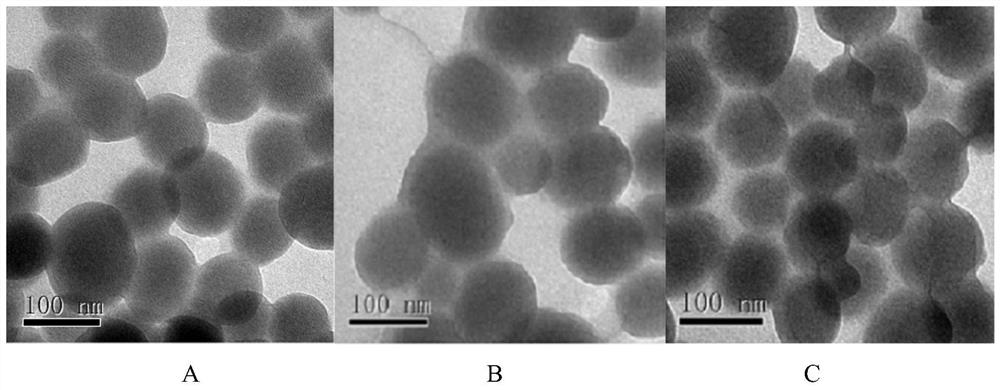
[0072] (1) Pre-synthesized MCM-41 type mesoporous silicon nanoparticles (synthesized according to the method in Colloids and Surfaces B: Biointerfaces, 2017, 155:41–50., with a particle size of 100nm and a pore size of 2-10nm) 60 After vacuum drying at ℃ for 24 hours, weigh 0.50 g and add it to 50 mL of anhydrous toluene, then add 0.20 mL of γ-aminopropyltriethoxysilane, stir evenly, raise the temperature to 110 ℃, and reflux for 24 hours under the protection of nitrogen. After the reaction was completed, it was filtered with suction, washed twice with toluene and once with ethanol, and dried in vacuum at 60° C. for 24 hours to obtain amino group-containing mesoporous silicon nanoparticles. Disperse 0.10 g of amino group-containing mesoporous silicon nanoparticles in acetone, add 0.20 g of succinic anhydride, stir and react at room temperature for 24 hours, and centrifuge to obtain carboxyl group-containing mesoporous silicon nanoparticles. Disperse 0.10g of carboxyl-containin...

Embodiment 2

[0078]The difference between embodiment 2 and embodiment 1 mainly lies in: this implementation step (4) and (5) replace the aminopolyethylene glycol monomethyl succinate of embodiment 1 with monomethoxy polyethylene glycol succinimide succinate Ether, so that the surface-coupled PEG can be broken into a stable covalent bond under slightly acidic conditions.

[0079] (1) Pre-synthesized MCM-41 type mesoporous silicon nanoparticles (Colloids and Surfaces B: Biointerfaces, 2017, 155:41–50.) (particle size 100nm, pore size 2-10nm) were vacuum-dried at 60°C for 24h , weighed 0.50g and added to 50mL of anhydrous toluene, then added 0.20mL of γ-aminopropyltriethoxysilane, stirred evenly, raised the temperature to 110°C, and refluxed for 24h under nitrogen protection. After the reaction was completed, it was filtered with suction, washed twice with toluene and once with ethanol, and dried in vacuum at 60° C. for 24 hours to obtain amino group-containing mesoporous silicon nanoparticle...

Embodiment 3

[0084] The difference between Example 3 and Example 1 mainly lies in that in this implementation step (3), N,N-diethylacrylamide is substituted for the N,N-dimethylacrylamide in Example 1.

[0085] (1) Vacuum-dry the pre-synthesized MCM-41 type mesoporous silicon nanoparticles at 60°C for 24 hours, weigh 0.50 g and add it to 50 mL of anhydrous toluene, then add 0.20 mL of γ-aminopropyltriethoxysilane, and stir evenly. The temperature was raised to 110° C., and the reaction was refluxed for 24 hours under the protection of nitrogen. After the reaction was completed, it was filtered with suction, washed twice with toluene and once with ethanol, and dried in vacuum at 50° C. for 24 hours to obtain amino group-containing mesoporous silicon nanoparticles. Disperse 0.10 g of amino group-containing mesoporous silicon nanoparticles in acetone, add 0.20 g of succinic anhydride, stir and react at room temperature for 24 hours, and centrifuge to obtain carboxyl group-containing mesoporou...


PUM

PropertyMeasurementUnit
particle sizeaaaaaaaaaa
sizeaaaaaaaaaa

Abstract

The invention discloses a tumor-specific cleavable PEG nanoparticles and a preparation method and application thereof. The invention modifies the surface of mesoporous silicon nanoparticles with cationic polymer layer containing amino group and disulfide bond, adds PEG containing benzaldehyde group at the end group, introduces PEG shell layer on the surface of the nanoparticles, and obtains tumor-specific cleavable PEG nanoparticles, wherein the surface of the mesoporous silicon nanoparticles is modified with cationic polymer layer containing amino group and disulfide bond, and then adds PEG containing benzaldehyde group at the end group. The carrier has the characteristics of PEG molecule shedding off in tumor microenvironment, responding to the redox of intracellular glutathione, strongdrug loading capacity, long blood circulation time, targeted drug delivery and other advantages, and is suitable for the preparation of anti-tumor drug carrier.

Description

technical field [0001] The invention belongs to the field of drug carriers, and in particular relates to a tumor-specific cleavable PEGylated nanoparticle, a preparation method and application. Background technique [0002] In recent years, the excellent performance of nanotechnology in drug delivery systems has attracted widespread attention, and it has achieved remarkable results in tumor treatment, showing great advantages and potential. Nano-drug carriers are loaded with drugs through physical methods, have a high drug loading capacity, can well maintain the activity of drugs, and can improve the targeted delivery ability and pharmacodynamic level of drugs to tumor tissues by strengthening the penetration and retention effects. Realize fixed-point, timed and quantitative release of drugs, not only reduce the toxic and side effects of drugs on normal tissues, but also give full play to the curative effect of drugs, so as to achieve the purpose of cure. [0003] Mesoporou...

Claims


Application Information

Patent Timeline
Patent Type & Authority Patents(China)
IPC IPC(8): A61K9/51A61K47/10A61K47/04A61K31/704A61P35/00
CPCA61K9/5115A61K9/5146A61K31/704A61P35/00
Inventor 王冠海林坚涛张大威于海兵
Owner GUANGDONG MEDICAL UNIV
Why Eureka
Try Eureka