Degradable controllable bone tissue engineering scaffold based on 3D printing and preparation method thereof
A technology of 3D printing and porous scaffolds, applied in tissue regeneration, medical science, prostheses, etc., can solve problems such as difficult to prepare pores smaller than 100µm and control surface nanoscale topological structure, so as to promote bone repair and reconstruction, biological The effect of good compatibility and good bone repair effect
- Summary
- Abstract
- Description
- Claims
- Application Information
AI Technical Summary
Problems solved by technology
Method used
Image
Examples
preparation example Construction
[0028] A method for preparing a 3D printed multi-level micro-nano structure controllable bioactive ceramic support, comprising the following steps:
[0029] 1. Prepare a bioactive ceramic scaffold with first-level macroscopic pores, and the specific preparation route is as follows:
[0030] (1) Mix calcium phosphate powder with bio-binder polyvinyl alcohol (PVB, Mn=70000-90000) and ethanol (Sigma) at a mass ratio of 3.6:32:32, and mix thoroughly to obtain calcium phosphate-based bioactive ceramics Print ink.
[0031] (2) The ceramic ink prepared above is used to prepare the target model by three-dimensional inkjet printing technology (3DP). During the printing process, the print head accumulates the target model layer by layer according to the set path, the selected print head diameter is 0.4mm, and the printing speed is 5-20mm / s. The printed model can be reversely reconstructed using 3D software based on the patient's bone defect data, and the internal porous structure of t...
Embodiment 1
[0039] (1) Configure calcium phosphate bioactive ceramic printing ink;
[0040] Thoroughly mix hydroxyapatite (HA) and tricalcium phosphate powder (TCP), wherein the mass ratio of hydroxyapatite (HA) to tricalcium phosphate (TCP) is 6:4, and the calcium phosphate powder and biological The binder polyvinyl alcohol (PVB, Mn=80000) and ethanol (Sigma) were fully mixed according to the mass ratio of 3.6:32:32 to prepare the calcium phosphate bioactive ceramic printing ink.
[0041] (2) Three-dimensional inkjet printing technology (3DP) is used to prepare a macroporous bioceramic body of the first level; the size of the first-level macroscopic macropores is designed by a three-dimensional design software. The nozzle diameter (d) is 200 μm, the slice thickness (h) is 160 μm for slice layering, and the printing speed is 300 mm / min for slurry extrusion 3D printing.
[0042] (3) Put the printed embryo body in step 2 into an oven to remove the solvent and sinter it into porcelain. The ...
Embodiment 2
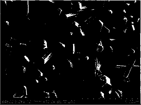
[0046] Calcium phosphate with a mass ratio of hydroxyapatite (HA) to tricalcium phosphate (TCP) of 3:7 was selected as the printing material. According to the method steps of Example 1, firstly, the first-level macro-scale macropore design 3D printing is carried out, and then the dissolution and recrystallization is carried out to carry out the second-level micro-nano scale surface topology regulation. The rest of the parameter selection and preparation process are the same as in Example 1. The advantage is that this embodiment adjusts the composition ratio of the raw materials and increases the ratio of tricalcium phosphate, which is more conducive to the re-dissolution and recrystallization of the material. Finally, an orthogonal porous bioceramic with a macro-scale macropore of 300 μm and a porosity of 70% was obtained. At the same time, the bioceramic had a micro-nano-scale topology on the surface of the second-level three-dimensional flaky whiskers, and the length of the f...
PUM
| Property | Measurement | Unit |
|---|---|---|
| Thickness | aaaaa | aaaaa |
| Length | aaaaa | aaaaa |
| Diameter | aaaaa | aaaaa |
Abstract
Description
Claims
Application Information
 - R&D
- Intellectual Property
- Life Sciences
- Materials
- Tech Scout
- Unparalleled Data Quality
- Higher Quality Content
- 60% Fewer Hallucinations
Browse by: Latest US Patents, China's latest patents, Technical Efficacy Thesaurus, Application Domain, Technology Topic, Popular Technical Reports.
© 2025 PatSnap. All rights reserved.Legal|Privacy policy|Modern Slavery Act Transparency Statement|Sitemap|About US| Contact US: help@patsnap.com


