Preparation method of magnetic resonance contrast agent targeting blood-brain barrier and glioma

A magnetic resonance contrast agent and mass ratio technology, which is applied in the direction of nuclear magnetic resonance/magnetic resonance imaging contrast agents, preparations for in vivo tests, medical preparations with non-active ingredients, etc., can solve the problem of lack of targeting and difficult Across the BBB, the body has potential toxicity and other problems, to achieve the effect of easy crossing the blood-brain barrier, simple and easy preparation process, and uniform particle size

Inactive Publication Date: 2019-03-01
AFFILIATED HUSN HOSPITAL OF FUDAN UNIV
View PDF7 Cites 7 Cited by
  • Summary
  • Abstract
  • Description
  • Claims
  • Application Information

AI Technical Summary

Problems solved by technology

[0004] The purpose of the present invention is to provide a method for preparing a magnetic resonance contrast agent targeting the blood-brain barrier and glioma; by preparing a kind of ultra-small superparamagnetic iron oxide nanoparticles (3 ~ 4nm), as MR positive contrast Construct a dual-targeting strategy that can target brain capillary endothelial cells and glioma cells at the same time, so as to solve the lack of targeting, difficulty crossing the BBB and harm to the body existing in the diagnosis of glioma. Potential toxicity and other issues

Method used


Image

  • Preparation method of magnetic resonance contrast agent targeting blood-brain barrier and glioma
  • Preparation method of magnetic resonance contrast agent targeting blood-brain barrier and glioma
  • Preparation method of magnetic resonance contrast agent targeting blood-brain barrier and glioma

Examples


Embodiment 1

[0038] (1) Preparation of ferric oxide nanoparticles coated with oleic acid: 2.7g ferric chloride hexahydrate was added in a 100ml round bottom flask, then 9.125g sodium oleate, 20ml ethanol, 15ml distilled water and 35ml n-hexane were added successively Alkanes were heated at 70°C for 4h in a magnetic stirrer while maintaining 700 rpm. Cool to room temperature after the reaction is complete. Wash the reacted liquid three times with distilled water and dry it overnight, leaving the waxy liquid that is ferric oleate; take out 1.8g of ferric oleate and add it to a three-necked flask, add 3.22g of oleyl alcohol and 10g of diphenyl ether. Under protection, heat the reaction system to 90°C for 2 hours to remove water and air in the solution, then continue to heat up to 200°C, keep for 30 minutes, precipitate with ethanol, dissolve and centrifuge with n-hexane to obtain black precipitates, namely iron ferric oxide nanoparticles, Dissolve in chloroform and save for later use.

[00...

Embodiment 2

[0044] 1: MR imaging effect:

[0045] 1.1 Experimental materials and instruments:

[0046] ANG / PEG‐USPIONs hydrophilic nanoparticles prepared in Example 1.

[0047] MR imaging detection instrument model: DiscoveryMR750, GE Medical System, 3.0T

[0048] 1.2. Experimental animals: Blab / c nude mice, with an average body weight of 20 g, were purchased from the animal room of Fudan University School of Medicine.

[0049] 1.3. Orthotopic glioma nude mouse model: U87MG cells (5×10 5 U87MG (dispersed in 5ul PBS) was implanted into the brain of nude mice with a stereotaxic instrument, and it was available after 14‐18 days.

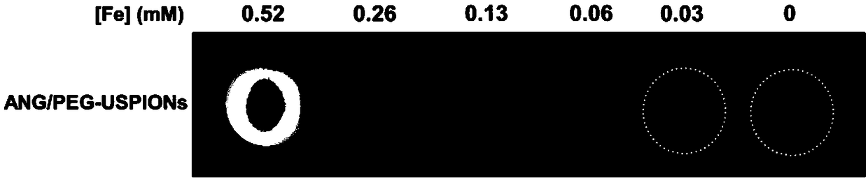
[0050] 1.4. Experimental method: Nude mice with glioma were anesthetized with chloral hydrate, injected contrast agent (5 mg Fe / ml) through the tail vein, and observed the effect of MRI.

[0051] 1.5. Experimental results: image 3 It is the in vitro MR imaging result of ANG / PEG-USPIONs hydrophilic nanoparticles, Figure 4 It can be seen that the prepared hyd...


PUM

PropertyMeasurementUnit
particle diameteraaaaaaaaaa
diameteraaaaaaaaaa
molecular weightaaaaaaaaaa

Abstract

The invention discloses a preparation method of a magnetic resonance contrast agent targeting blood-brain barrier and glioma. The method comprises the following steps of dispersing ferric chloride hexahydrate and sodium oleate in a mixture of ethanol, water and n-hexane to obtain precursor iron oleate; dissolving the precursor in a mixed solution of oleoyl alcohol and diphenyl ether, and stirringthe mixture at about 110 DEG C under nitrogen protection; rising the temperature to about 200 DEG C under nitrogen protection, and carrying out reflux to obtain an oil acid-coated ferroferric oxide nanoparticles; and dispersing the nanoparticles in trichloromethane, adding excess DSPE-PEG-Maleimide for hydrophilic modification, dispersing the modified ferroferric oxide nanoparticles in PBS, and finally grafting specific glioma-targeted molecules Angiopep-2. The magnetic resonance contrast agent can efficiently cross the blood-brain barrier, and can be used as a novel type of magnetic resonanceimaging positive contrast agent for early diagnosis and boundary definition of glioma.

Description

technical field [0001] The invention belongs to the technical field of molecular imaging, and relates to a preparation method of a magnetic resonance contrast agent targeting the blood-brain barrier and brain glioma. Background technique [0002] Glioma is the most common central nervous system tumor, accounting for about 35‐45% of the incidence of intracranial tumors. At present, the main treatment for glioma is surgical resection. However, due to its infiltrative growth pattern and the difficulty of accurately delineating the boundaries of glioma with current examination methods, surgery cannot achieve complete removal of tumor tissue. Magnetic resonance imaging (Magnetic Resonance Imaging, MRI) has the advantages of high tissue resolution, multi-parameter, multi-sequence imaging, and no radiation. It is the main imaging method for clinical detection of central nervous system diseases. Therefore, the preparation of MR probes that can specifically target gliomas and the ac...

Claims


Application Information

Patent Timeline
Patent Type & Authority Applications(China)
IPC IPC(8): A61K49/14A61K49/08A61K47/24A61K47/62
CPCA61K49/085A61K49/14
Inventor 张军杜成娟王剑虹陈雨耿道颖
Owner AFFILIATED HUSN HOSPITAL OF FUDAN UNIV
Why Eureka
Try Eureka