A method for display intraosseous vein-artery blood vessel

An arterial blood vessel and display method technology, which is applied in the fields of healthcare informatics, instruments, computing, etc., can solve the problems of affecting data analysis, inability to measure the diameter of blood vessels, and cumbersome operations, so as to simplify the experimental process, shorten the research cycle, and improve the efficiency of research. Precise results

Active Publication Date: 2019-03-12
AFFILIATED ZHONGSHAN HOSPITAL OF DALIAN UNIV
View PDF3 Cites 1 Cited by
  • Summary
  • Abstract
  • Description
  • Claims
  • Application Information

AI Technical Summary

Problems solved by technology

[0003] According to the researched data, there are currently: (1) Gelatin lead oxide perfusion technology combined with clinical CT to display blood vessels is limited by resolution, and can only reconstruct and quantitatively study the structure of larger diameter blood vessels, and the resolution of clinical CT is still not enough. for femoral head intraosseous veins for vascular research
(2) Superselective DSA angiography of the hip can display images of venous return to the femoral head, but it cannot be quantified, and the resolution is low, which is not enough to observe and study the three-dimensional distribution of small blood vessels in the femoral head
After systemic injection of contrast agent, the blood supply status of the femoral head can be evaluated by quantifying the signal changes, but the three-dimensional distribution of the femoral head veins and the quantitative study of the veins cannot be carried out
(4) There are no technical methods and research reports on the three-dimensional distribution of femoral head and vein vessels
(5) Research reports showing the three-dimensional distribution of femoral head vein-artery and the spatial relationship between femoral head vein and artery at the same time have not been reported at home and abroad
[0005] Defects or problems in the prior art: (1) The femoral head retinaculum vein is thin and difficult to find
(2) It is easy to retain thrombus in the venous pipeline system to block the venous vascular system
(3) There may be valve perfusion fluid in the venous pipeline system that can only flow centripetally, and it is difficult to succeed in venous perfusion
(4) Lead oxide is poisonous, the atomic number of lead is large, and the ray attenuation coefficient is high. After clinical CT scanning, there are glitches and artifacts after image data reconstruction, which affects data analysis.
(5) The maximum resolution of clinical CT is 0.65mm, and the diameter of intraosseous vessels is mostly smaller than this resolution, so intraosseous vessels cannot be accurately displayed
Traditional research methods cannot perform quantitative calculations such as vessel diameter measurement, length measurement, length density calculation, and volume density calculation.
(7) Since the problem of vascular perfusion display of the femoral head vein has not been solved, the co-display of the femoral head vein-artery is also an unsolved worldwide research problem
Defects in technical methods: ①. Destruction of the integrity of intraosseous blood vessels
② No matter how the slices of the femoral head are placed or overlapped, the image obtained by X-ray photography is always a two-dimensional image, which is not convenient for three-dimensional observation, measurement and presentation
③, the production process is time-consuming and cumbersome to operate
④. At present, there is no reliable and exact method for the study of intraosseous vein-artery vascular perfusion, three-dimensional reconstruction and co-display

Method used


Image

  • A method for display intraosseous vein-artery blood vessel
  • A method for display intraosseous vein-artery blood vessel
  • A method for display intraosseous vein-artery blood vessel

Examples


Embodiment 1

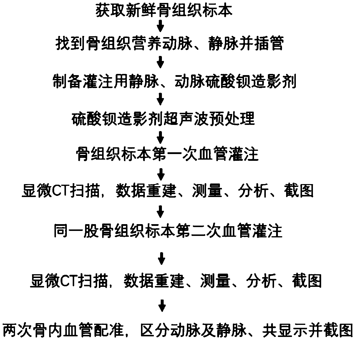
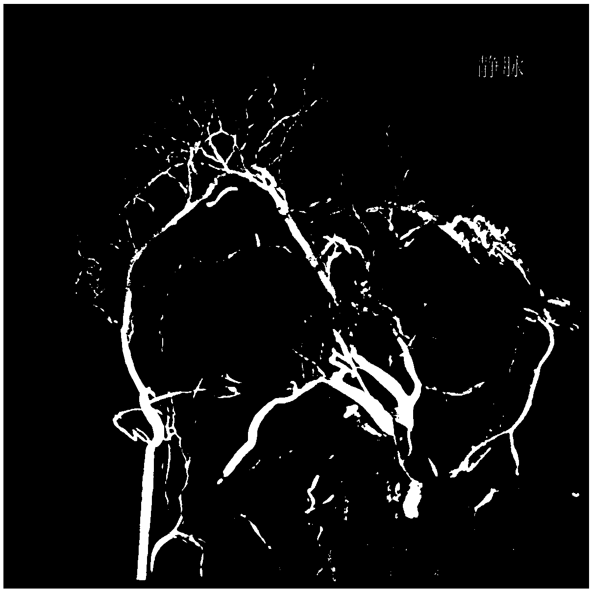
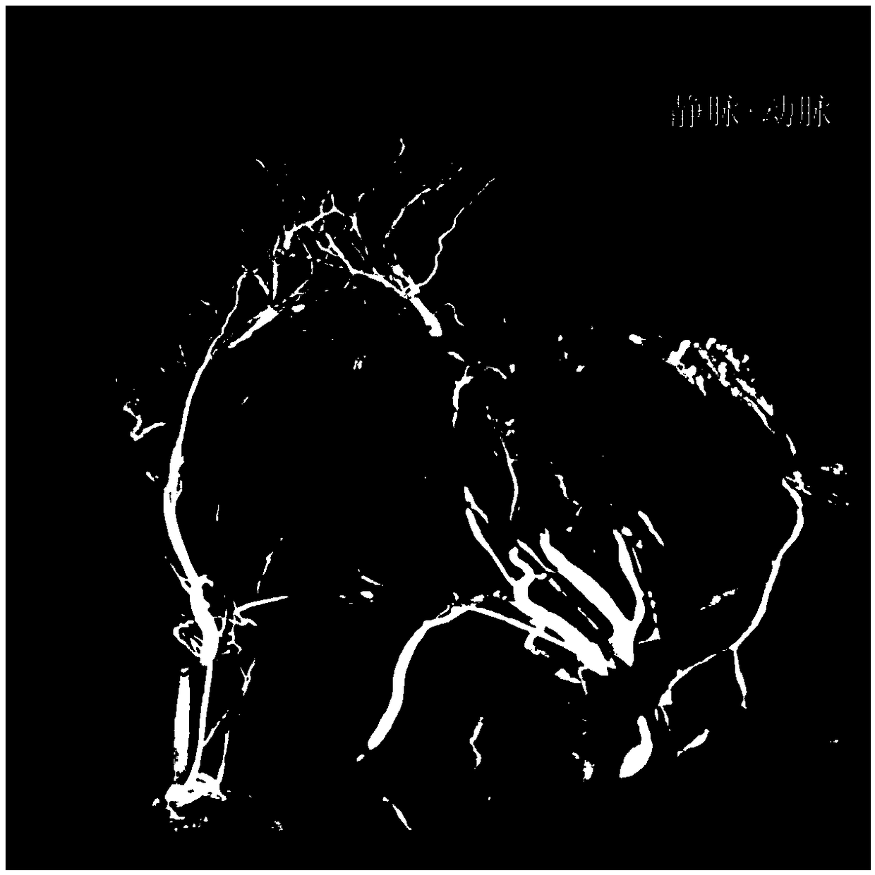
[0059] Inferior retinal vein and arterial cannulation, retrograde perfusion of femoral head vein with gelatin barium sulfate contrast medium, antegrade perfusion of femoral head artery with gelatin barium sulfate contrast medium

[0060] (1) Use fresh pig femoral head specimens.

[0061] (2) Intubation of retinal veins and arteries: magnify 10 times under a microscope, use microscopic instruments to perform micromanipulation, and free retinaculum vessels. At the back of the femoral head and neck, look for the retinaculum vein under the femoral head, and use the back of the scalpel to squeeze the soft tissue of the femoral head and neck from the distal end to the proximal segment. At this time, it can be seen that there is venous blood in the venous lumen and it is filled with thin of the venous lumen. A 6# flat needle (outer diameter 0.6mm) was selected, the front of the needle was smooth, and the back was rough. The perfusion needle was intubated, ligated and fixed. Posteri...

Embodiment 2

[0073] Inferior retinal vein and arterial cannulation, carboxymethylcellulose barium sulfate contrast medium perfusion retrograde femoral head vein, carboxymethylcellulose barium sulfate contrast medium antegrade perfusion femoral head artery

[0074] (1) Use fresh pig femoral head specimens.

[0075] (2) Intubation of retinal veins and arteries: magnify 10 times under a microscope, use microscopic instruments to perform micromanipulation, and free retinaculum vessels. At the back of the femoral head and neck, look for the retinaculum vein under the femoral head, and use the back of the scalpel to squeeze the soft tissue of the femoral head and neck from the distal end to the proximal segment. At this time, it can be seen that there is venous blood in the venous lumen and it is filled with thin of the venous lumen. A 6# flat needle (outer diameter 0.6mm) was selected, the front of the needle was smooth, and the back was rough. The perfusion needle was intubated, ligated and f...

Embodiment 3

[0082] Inferior retinal vein and arterial cannulation, carboxymethylcellulose barium sulfate contrast medium perfusion retrograde femoral head vein, carboxymethylcellulose barium sulfate contrast medium antegrade perfusion femoral head artery

[0083] (1) Use fresh pig femoral head specimens.

[0084] (2) Intubation of retinal veins and arteries: magnify 10 times under a microscope, use microscopic instruments to perform micromanipulation, and free retinaculum vessels. At the back of the femoral head and neck, look for the retinaculum vein under the femoral head, and use the back of the scalpel to squeeze the soft tissue of the femoral head and neck from the distal end to the proximal segment. At this time, it can be seen that there is venous blood in the venous lumen and it is filled with thin of the venous lumen. A 6# flat needle (outer diameter 0.6mm) was selected, the front of the needle was smooth, and the back was rough. The perfusion needle was intubated, ligated and f...


PUM


Abstract

The invention discloses a method for display intraosseous vein-arterial blood vessel. The method comprises that follow steps: (1) performing first blood vessel perfusion on bone tissue specimen with perfusion liquid A or perfusion liquid B, performing microCT scan and first three-dimensional reconstruction of intraosseous blood vessel data to obtain first intraosseous blood vessel data; (2) Perfusion solution A or perfusion solution B was used to perfusion the second blood vessel of bone tissue, and the second three-dimensional reconstruction of intraosseous blood vessel data was performed bymicro-CT scanning to obtain the second blood vessel data; (3) Register the first intraosseous blood vessel data and the second intraosseous blood vessel data to obtain the femoral artery blood vesselsystem and femoral vein blood vessel system. The invention is the first, simplest and most time-saving method, the research result is accurate, and can quantify the blood vessel diameter, blood vessellength, blood vessel volume, blood vessel length density, volume density, bone trabecular parameters, and is interrelated with bone mineral content in bone tissue and bone trabecular structure parameters.

Description

technical field [0001] The present invention relates to osteonecrosis (such as: femoral head, humeral head, femoral condyle, patella, talus, scaphoid, metatarsal, or lunate osteonecrosis, etc.), osteoporosis, anatomical pipeline research techniques and methods for bone circulation research and perfusion Material. Used to display and study intraosseous vein-artery co-distribution, construction. And it can quantitatively compare and study the intraosseous vein-artery status in different pathophysiological states. To study the structure, three-dimensional distribution, quantification and construction of veins and arteries in the pathophysiological changes of femoral neck fractures, avascular osteonecrosis of the femoral head, osteoarthritis, and osteoporosis. Quantitative research on the making of vein-artery teaching model of intraosseous vascular structure and the construction of three-dimensional finite element hemodynamic model of vein-artery. This technique can be applied...

Claims


Application Information

Patent Timeline
Patent Type & Authority Applications(China)
IPC IPC(8): G06T15/00G16H30/40
CPCG06T15/005G16H30/40
Inventor 赵德伟邱兴徐达传欧阳钧刘继峰
Owner AFFILIATED ZHONGSHAN HOSPITAL OF DALIAN UNIV
Why Eureka
Try Eureka